

Role of the hippocampus, the bed nucleus of the stria terminalis, and the amygdala in the excitatory effect of corticotropin-releasing hormone on the acoustic startle reflex
- PMID: 9236251
- PMCID: PMC6568348
- DOI: 10.1523/JNEUROSCI.17-16-06434.1997
Role of the hippocampus, the bed nucleus of the stria terminalis, and the amygdala in the excitatory effect of corticotropin-releasing hormone on the acoustic startle reflex
Abstract
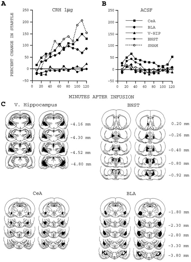
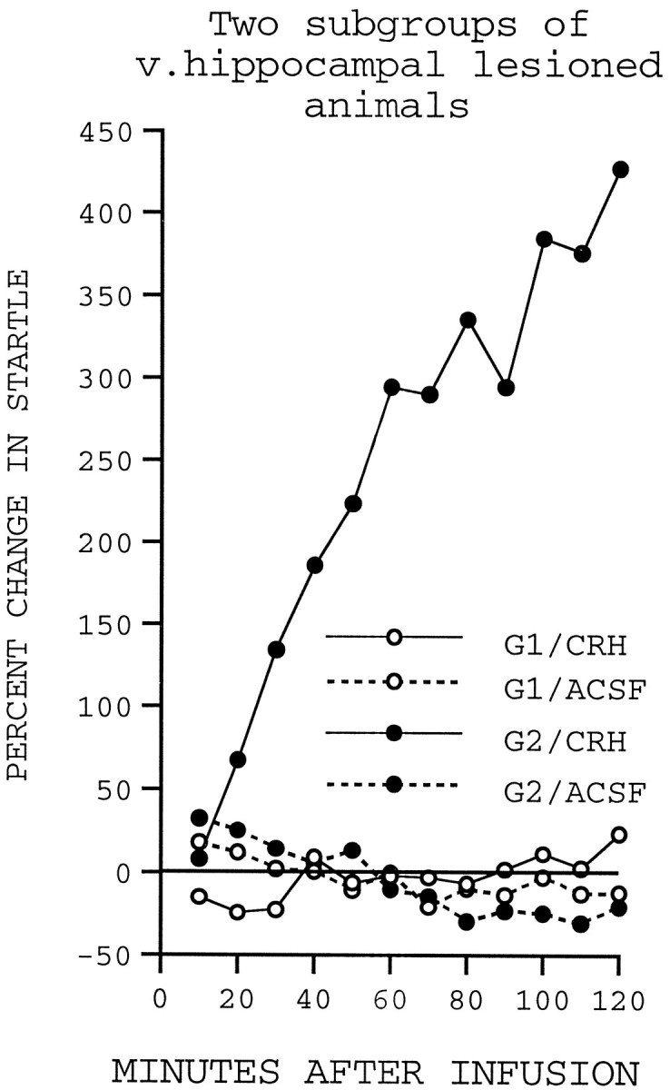
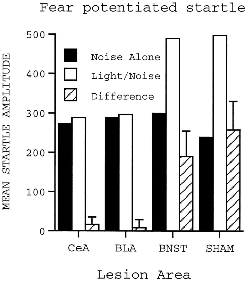
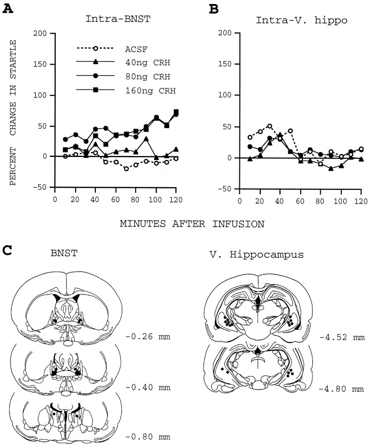
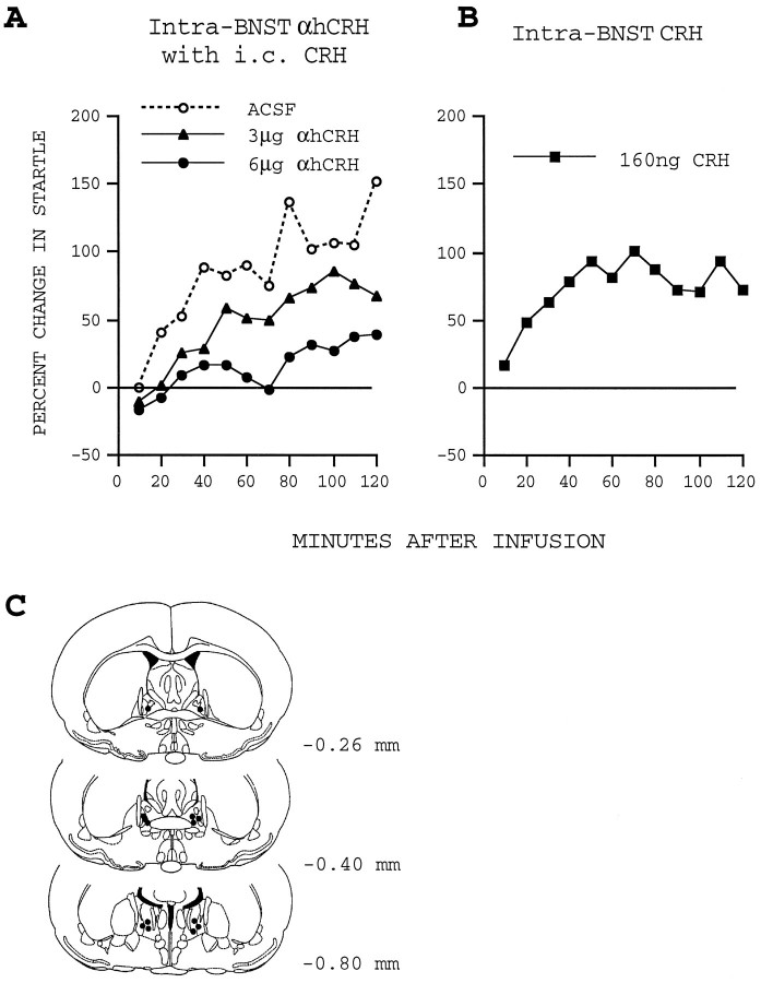
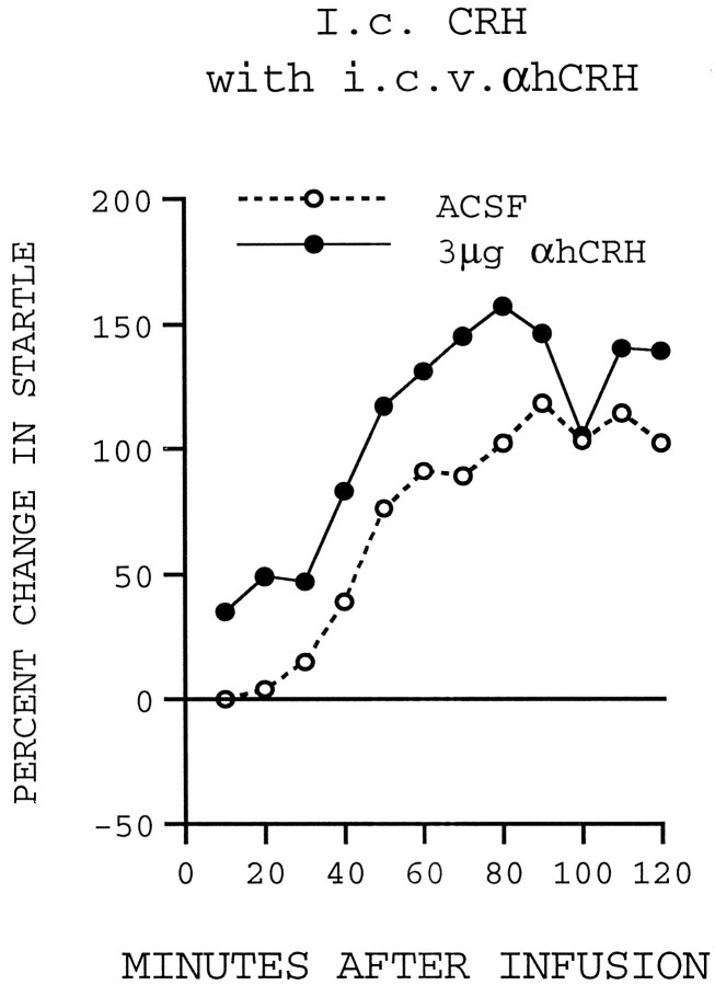
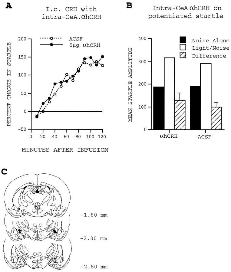
Previously, we demonstrated that transection of the fimbria/fornix blocked the excitatory effect of corticotropin-releasing hormone (CRH) on startle (CRH-enhanced startle), suggesting that the hippocampus and its efferent target areas that communicate via the fimbria may be critically involved in CRH-enhanced startle. The bed nucleus of the stria terminalis (BNST) receives direct projections from the ventral hippocampus via the fimbria/fornix. Therefore, the role of the ventral hippocampus, the BNST, and the amygdala in CRH-enhanced startle was investigated. NMDA lesions of the BNST completely blocked CRH-enhanced startle, whereas chemical lesions of the ventral hippocampus and the amygdala failed to block CRH-enhanced startle. However, the same amygdala-lesioned animals showed a complete blockade of fear-potentiated startle, a conditioned fear response sensitive to manipulations of the amygdala. In contrast, BNST-lesioned rats had normal fear-potentiated startle. This indicates a double dissociation between the BNST and the amygdala in two different paradigms that enhance startle amplitude. Microinfusions of CRH into the BNST, but not into the ventral hippocampus, mimicked intracerebroventricular CRH effects. Furthermore, infusion of a CRH antagonist into the BNST blocked CRH-enhanced startle in a dose-dependent manner. Control studies showed that this blockade did not result from either leakage of the antagonist into the ventricular system or a local anesthetic effect caused by infusion of the antagonist into the BNST. The present studies strongly suggest that CRH in the CSF can activate the BNST, which could lead to activation of brainstem and hypothalamic BNST target areas involved in anxiety and stress responses.
Figures
Similar articles
-
Role of the septum in the excitatory effect of corticotropin-releasing hormone on the acoustic startle reflex.J Neurosci. 1997 Aug 15;17(16):6424-33. doi: 10.1523/JNEUROSCI.17-16-06424.1997. J Neurosci. 1997. PMID: 9236250 Free PMC article.
-
Evidence that preimmunization with a heat-killed preparation of Mycobacterium vaccae reduces corticotropin-releasing hormone mRNA expression in the extended amygdala in a fear-potentiated startle paradigm.Brain Behav Immun. 2019 Mar;77:127-140. doi: 10.1016/j.bbi.2018.12.015. Epub 2018 Dec 28. Brain Behav Immun. 2019. PMID: 30597198
-
Modulation of the acoustic startle reflex by infusion of corticotropin-releasing hormone into the nucleus reticularis pontis caudalis.Brain Res. 1998 Jan 26;782(1-2):318-23. doi: 10.1016/s0006-8993(97)01309-7. Brain Res. 1998. PMID: 9519280
-
Role of the extended amygdala in short-duration versus sustained fear: a tribute to Dr. Lennart Heimer.Brain Struct Funct. 2008 Sep;213(1-2):29-42. doi: 10.1007/s00429-008-0183-3. Epub 2008 Jun 5. Brain Struct Funct. 2008. PMID: 18528706 Review.
-
Role of the bed nucleus of the stria terminalis versus the amygdala in fear, stress, and anxiety.Eur J Pharmacol. 2003 Feb 28;463(1-3):199-216. doi: 10.1016/s0014-2999(03)01282-2. Eur J Pharmacol. 2003. PMID: 12600711 Review.
Cited by
-
Chronic administration of the triazolobenzodiazepine alprazolam produces opposite effects on corticotropin-releasing factor and urocortin neuronal systems.J Neurosci. 2000 Feb 1;20(3):1240-8. doi: 10.1523/JNEUROSCI.20-03-01240.2000. J Neurosci. 2000. PMID: 10648728 Free PMC article.
-
Deletion of CRH From GABAergic Forebrain Neurons Promotes Stress Resilience and Dampens Stress-Induced Changes in Neuronal Activity.Front Neurosci. 2019 Sep 20;13:986. doi: 10.3389/fnins.2019.00986. eCollection 2019. Front Neurosci. 2019. PMID: 31619956 Free PMC article.
-
Ancestral Stress Alters Lifetime Mental Health Trajectories and Cortical Neuromorphology via Epigenetic Regulation.Sci Rep. 2019 Apr 23;9(1):6389. doi: 10.1038/s41598-019-42691-z. Sci Rep. 2019. PMID: 31011159 Free PMC article.
-
Inhibition of corticotropin releasing factor expression in the central nucleus of the amygdala attenuates stress-induced behavioral and endocrine responses.Front Neurosci. 2013 Oct 29;7:195. doi: 10.3389/fnins.2013.00195. eCollection 2013. Front Neurosci. 2013. PMID: 24194694 Free PMC article.
-
Early-Life Stress Impairs Perception and Neural Encoding of Rapid Signals in the Auditory Pathway.J Neurosci. 2023 May 3;43(18):3232-3244. doi: 10.1523/JNEUROSCI.1787-22.2023. Epub 2023 Mar 27. J Neurosci. 2023. PMID: 36973014 Free PMC article.
References
-
- Alheid GF, de Olmos JS, Beltramino CA. Amygdala and extended amygdala. In: Paxinos G, editor. The rat nervous system. Academic; San Diego: 1995. pp. 495–578.
-
- Amaral DG, Witter MP. Hippocampal formation. In: Paxinos G, editor. The rat nervous system. Academic; San Diego: 1995. pp. 443–493.
-
- Arató M, Bánki C, Bissette G, Nemeroff CB. Elevated CSF CRH in suicide victims. Biol Psychiatry. 1989;25:255–359. - PubMed
-
- Arnold FJL, Bueno MD, Shiers H, Hancock DC, Evan GI, Herbert J. Expression of c-fos in regions of the basal limbic forebrain following intracerebroventricular corticotropin-releasing factor in unstressed or stressed male rats. Neuroscience. 1992;51:377–390. - PubMed
-
- Bánki CM, Karmacsi L, Bissette G, Nemeroff CB. Cerebrospinal fluid neuropeptides in mood disorder and dementia. J Affect Disord. 1992a;25:39–45. - PubMed
Publication types
MeSH terms
Substances
Grants and funding
LinkOut - more resources
Full Text Sources
Other Literature Sources