

Regeneration of a central synapse restores nonassociative learning
- PMID: 9236255
- PMCID: PMC6568365
- DOI: 10.1523/JNEUROSCI.17-16-06478.1997
Regeneration of a central synapse restores nonassociative learning
Abstract
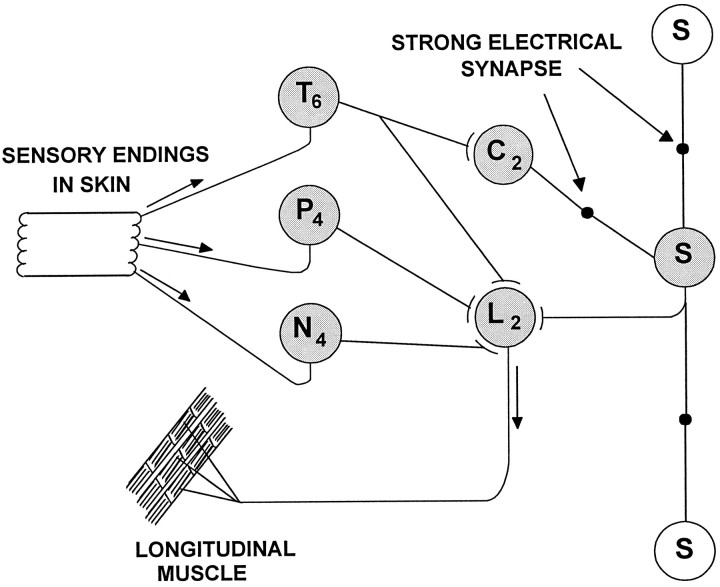
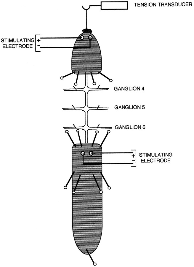
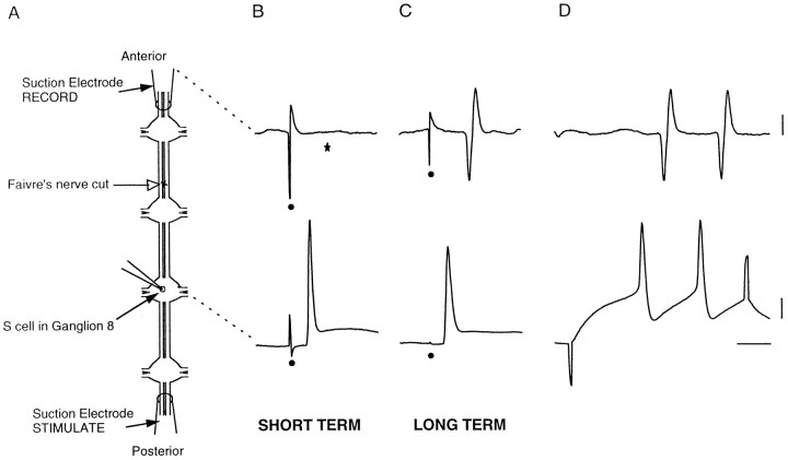
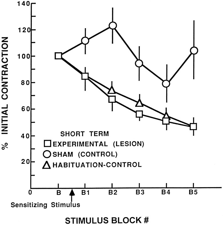
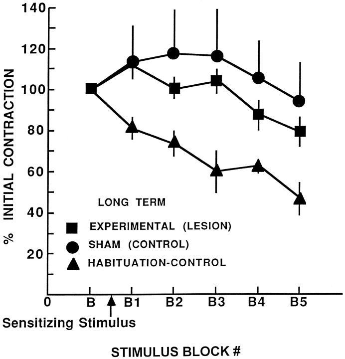
Sensitization is a form of nonassociative learning in which a strong or noxious stimulus persistently enhances the response produced by a weaker stimulus. In the leech Hirudo medicinalis, the S-interneuron is required for sensitization of the shortening response. A single S-cell axon was surgically separated from its sole synaptic partner, the neighboring S-cell. This consistently eliminated sensitization without impairing reflexive shortening itself, as measured in semi-intact specimens. Sensitization of the shortening reflex returned after 3 weeks when the severed axon grew and regenerated its specific electrical synapse within the nerve cord, as shown by restored conduction of impulses between S-cells. This confirms the essential role of one neuron, the S-cell, in sensitization, and it demonstrates that regeneration of the synapse between S-cells restores this example of nonassociative learning.
Figures
Similar articles
-
Repair and regeneration of functional synaptic connections: cellular and molecular interactions in the leech.Cell Mol Neurobiol. 2005 Mar;25(2):441-50. doi: 10.1007/s10571-005-3152-x. Cell Mol Neurobiol. 2005. PMID: 16047551 Free PMC article. Review.
-
Progressive recovery of learning during regeneration of a single synapse in the medicinal leech.J Comp Neurol. 2003 Feb 24;457(1):67-74. doi: 10.1002/cne.10530. J Comp Neurol. 2003. PMID: 12541325
-
Accurate synapse regeneration despite ablation of the distal axon segment.Eur J Neurosci. 1996 Jan;8(1):11-20. doi: 10.1111/j.1460-9568.1996.tb01163.x. Eur J Neurosci. 1996. PMID: 8713446
-
Lasting changes in a network of interneurons after synapse regeneration and delayed recovery of sensitization.Neuroscience. 2007 Dec 19;150(4):915-25. doi: 10.1016/j.neuroscience.2007.09.061. Epub 2007 Oct 5. Neuroscience. 2007. PMID: 18031937 Free PMC article.
-
Tinkering with successful synapse regeneration in the leech: adding insult to injury.J Exp Biol. 1987 Sep;132:207-21. doi: 10.1242/jeb.132.1.207. J Exp Biol. 1987. PMID: 3323400 Review.
Cited by
-
Serotonin mediates stress-like effects on responses to non-nociceptive stimuli in the medicinal leech Hirudo verbana.J Exp Biol. 2022 Jun 1;225(11):jeb243404. doi: 10.1242/jeb.243404. Epub 2022 Jun 9. J Exp Biol. 2022. PMID: 35510636 Free PMC article.
-
Repair and regeneration of functional synaptic connections: cellular and molecular interactions in the leech.Cell Mol Neurobiol. 2005 Mar;25(2):441-50. doi: 10.1007/s10571-005-3152-x. Cell Mol Neurobiol. 2005. PMID: 16047551 Free PMC article. Review.
-
Associative, bidirectional changes in neural signaling utilizing NMDA receptor- and endocannabinoid-dependent mechanisms.Learn Mem. 2011 Aug 15;18(9):545-53. doi: 10.1101/lm.2252511. Print 2011. Learn Mem. 2011. PMID: 21844187 Free PMC article.
-
Injury alters sensory, motor, and integrative elements underlying operant conditioning in the medicinal leech.PLoS One. 2025 Jun 12;20(6):e0326039. doi: 10.1371/journal.pone.0326039. eCollection 2025. PLoS One. 2025. PMID: 40504817 Free PMC article.
-
Endocannabinoid-dependent LTD in a nociceptive synapse requires activation of a presynaptic TRPV-like receptor.J Neurophysiol. 2010 Nov;104(5):2766-77. doi: 10.1152/jn.00491.2010. Epub 2010 Sep 8. J Neurophysiol. 2010. PMID: 20884761 Free PMC article.
References
-
- Bagnoli P, Brunelli M, Magni F, Pellegrino M. The neuron of the fast conducting system in Hirudo medicinalis: identification and synaptic connections with primary afferent neurons. Arch Ital Biol. 1975;113:21–43. - PubMed
-
- Byrne JH, Crow T. Examples of mechanistic analysis of learning and memory in invertebrates. In: Kesner R, Martinez J, editors. Learning and memory. Academic; New York: 1991. pp. 329–358.
-
- Calabrese RL, De Schutter E. Motor-pattern-generating networks in invertebrates: modeling our way toward understanding. Trends Neurosci. 1992;15:439–445. - PubMed
Publication types
MeSH terms
Grants and funding
LinkOut - more resources
Full Text Sources