. 1997 Aug 5;94(16):8405-10.
doi: 10.1073/pnas.94.16.8405.

Monitoring protein-protein interactions in intact eukaryotic cells by beta-galactosidase complementation

Affiliations

Monitoring protein-protein interactions in intact eukaryotic cells by beta-galactosidase complementation

F Rossi et al. Proc Natl Acad Sci U S A. .

Abstract

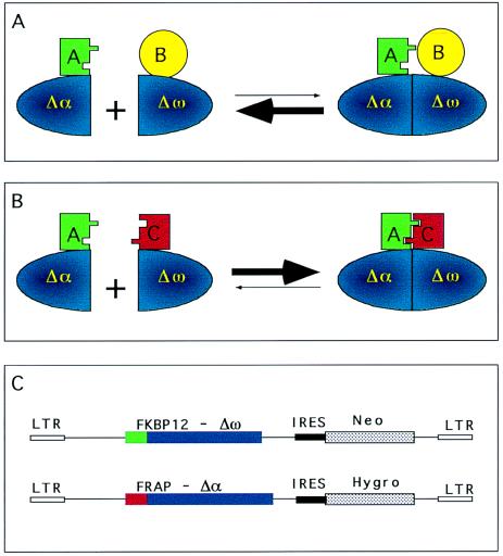
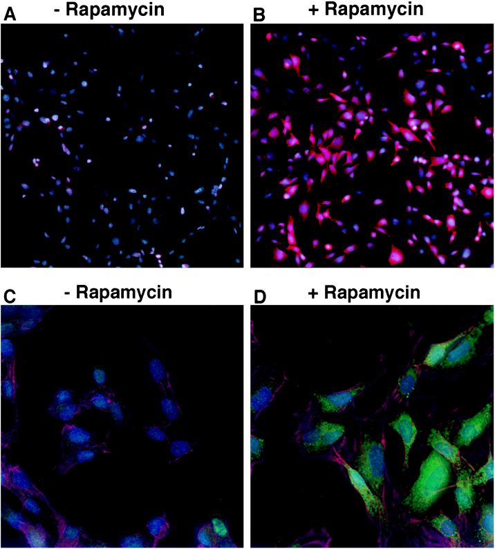
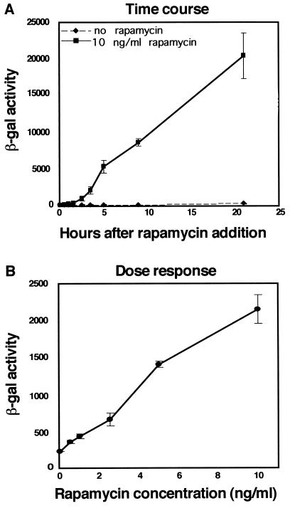
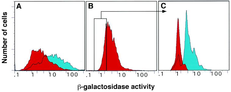
We present an approach for monitoring protein-protein interactions within intact eukaryotic cells, which should increase our understanding of the regulatory circuitry that controls the proliferation and differentiation of cells and how these processes go awry in disease states such as cancer. Chimeric proteins composed of proteins of interest fused to complementing beta-galactosidase (beta-gal) deletion mutants permit a novel analysis of protein complexes within cells. In this approach, the beta-gal activity resulting from the forced interaction of nonfunctional weakly complementing beta-gal peptides (Deltaalpha and Deltaomega) serves as a measure of the extent of interaction of the non-beta-gal portions of the chimeras. To test this application of lacZ intracistronic complementation, proteins that form a complex in the presence of rapamycin were used. These proteins, FRAP and FKBP12, were synthesized as fusion proteins with Deltaalpha and Deltaomega, respectively. Enzymatic beta-gal activity served to monitor the formation of the rapamycin-induced chimeric FRAP/FKBP12 protein complex in a time- and dose-dependent manner, as assessed by histochemical, biochemical, and fluorescence-activated cell sorting assays. This approach may prove to be a valuable adjunct to in vitro immunoprecipitation and crosslinking methods and in vivo yeast two-hybrid and fluorescence energy transfer systems. It may also allow a direct assessment of specific protein dimerization interactions in a biologically relevant context, localized in the cell compartments in which they occur, and in the milieu of competing proteins.


Figures

Figure 1
Figure 1
Experimental design. (A) When the Δα and Δω β-gal mutants are fused to proteins that do not dimerize, their association is not favored and β-gal activity is not detected. (B) When the Δα and Δω β-gal mutants are fused to proteins that can dimerize, the formation of active β-gal is favored. (C) Schematic representation of the FKBP12-Δω-Neo and the FRAP-Δα-Hygro constructs. IRES, internal ribosome entry sequence; LTR, long terminal repeat.
Figure 3
Figure 3
Histochemical assay of induction of β-gal activity upon chimera complementation. C2C12 cells expressing both FKBP12-Δω and FRAP-Δα were maintained overnight either in the absence (A and C) or the presence (B and D) of 10 ng/ml rapamycin. β-gal activity was visualized by fluorescence microscopy using Fluor-X-Gal as substrate. (A and B) Double-labeled samples showing Hoechst stained nuclei (blue) and β-gal activity using Fluor-X-Gal as substrate viewed with a rhodamine filter set (red). (C and D) Triple-labeled samples obtained by imaging with a DeltaVision microscope showing β-gal activity using Fluor-X-Gal as substrate (green), Hoechst stained nuclei (blue), and Cy5-labeled actin filaments (red) to visualize the contour of each cell at higher magnification.
Figure 2
Figure 2
Biochemical assay of induction of β-gal activity upon chimera complementation. (A) Kinetics of induction of β-gal activity upon treatment with rapamycin. Pure populations of C2C12 cells stably expressing both FKBP12-Δω and FRAP-Δα were plated in 96-well plates and 10 ng/ml rapamycin was added at time zero. Cells were then lysed at different time intervals thereafter, and the β-gal activity in the lysates was quantitated by chemiluminescence. (B) Dose response of β-gal activity upon rapamycin treatment. C2C12 cells expressing both FKBP12-Δω and FRAP-Δα were plated in 96-well plates and treated with different concentrations of rapamycin for 3.5 hr. β-gal activity is expressed as luminescence counts per second. Each point represents the average of six replicate samples. Error bars indicate standard deviations from the mean.
Figure 4
Figure 4
FACS analysis of induced β-gal activity upon chimera complementation. The red peaks represent the untreated samples and the blue peaks represent samples treated with 10 ng/ml rapamycin. (A) Induction of β-gal activity in a population of C2C12 cells expressing both FKBP12-Δω and FRAP-Δα after 90 min of rapamycin treatment. The majority of the cells respond to rapamycin treatment with an increase in β-gal activity. (B) Subpopulation of cells selected on the basis of low β-gal activity in uninduced conditions. (C) The same population was maintained overnight in the absence (red peak) or in the presence (blue peak) of rapamycin. The induced and uninduced populations yield essentially nonoverlapping peaks. The vertical axis represents relative cell number and the horizontal axis represents intensity of β-gal fluorescence on a logarithmic scale.

References

    1. Gu W, Schneider J W, Condorelli G, Kaushal S, Mahdavi V, Nadal-Ginard B. Cell. 1993;72:309–324. - PubMed
    1. Halevy O, Novitch B G, Spicer D B, Skapek S X, Rhee J, Hannon G J, Beach D, Lassar A B. Science. 1995;267:1018–1021. - PubMed
    1. Schneider J W, Gu W, Zhu L, Mahdavi V, Nadal-Ginard B. Science. 1994;264:1467–1471. - PubMed
    1. Bai C, Elledge S J. Methods Enzymol. 1996;273:331–347. - PubMed
    1. Fields S, Song O. Nature (London) 1989;340:245–246. - PubMed

Publication types

LinkOut - more resources