. 1997 Aug 5;94(16):8411-6.
doi: 10.1073/pnas.94.16.8411.

Three-dimensional structure of NADPH-cytochrome P450 reductase: prototype for FMN- and FAD-containing enzymes

Affiliations

Three-dimensional structure of NADPH-cytochrome P450 reductase: prototype for FMN- and FAD-containing enzymes

M Wang et al. Proc Natl Acad Sci U S A. .

Abstract

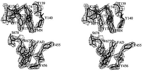
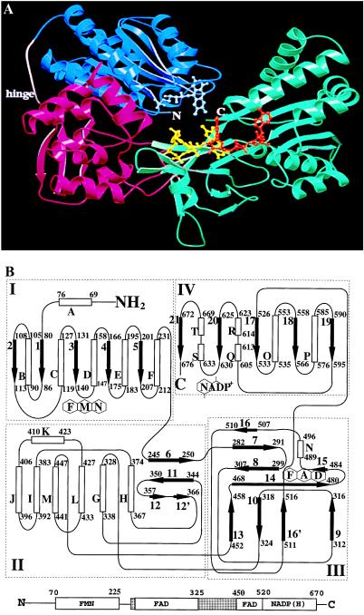
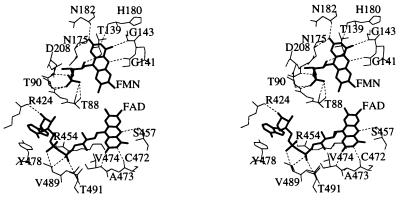
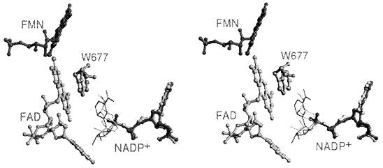
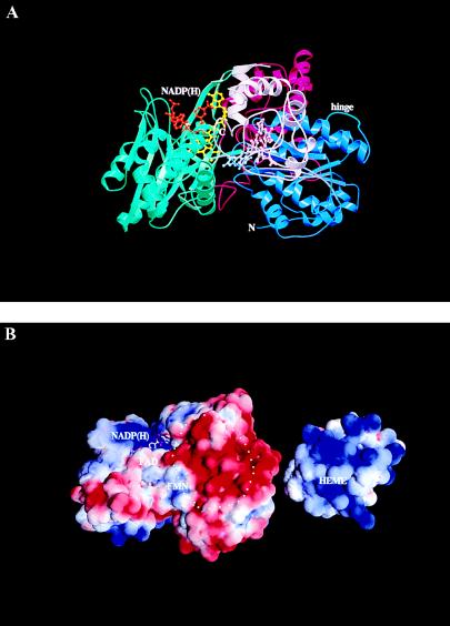
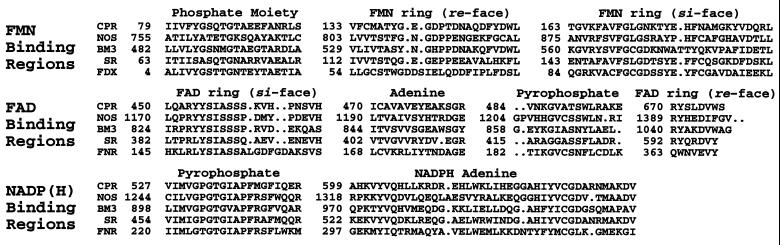
Microsomal NADPH-cytochrome P450 reductase (CPR) is one of only two mammalian enzymes known to contain both FAD and FMN, the other being nitric-oxide synthase. CPR is a membrane-bound protein and catalyzes electron transfer from NADPH to all known microsomal cytochromes P450. The structure of rat liver CPR, expressed in Escherichia coli and solubilized by limited trypsinolysis, has been determined by x-ray crystallography at 2.6 A resolution. The molecule is composed of four structural domains: (from the N- to C- termini) the FMN-binding domain, the connecting domain, and the FAD- and NADPH-binding domains. The FMN-binding domain is similar to the structure of flavodoxin, whereas the two C-terminal dinucleotide-binding domains are similar to those of ferredoxin-NADP+ reductase (FNR). The connecting domain, situated between the FMN-binding and FNR-like domains, is responsible for the relative orientation of the other domains, ensuring the proper alignment of the two flavins necessary for efficient electron transfer. The two flavin isoalloxazine rings are juxtaposed, with the closest distance between them being about 4 A. The bowl-shaped surface near the FMN-binding site is likely the docking site of cytochrome c and the physiological redox partners, including cytochromes P450 and b5 and heme oxygenase.


Figures

Figure 1
Figure 1
Stereoview of the electron density in the vicinity of the FAD and FMN of CPR. The 3|Fo| − 2|Fc| electron density map computed with 2.6 Å resolution data is contoured at 1.2σ level. Residues within close contact of the flavin ring are labeled. Four aromatic residues sandwich the flavin rings: Y178 and Y140 for FMN and Y456 and W677 for FAD.
Figure 2
Figure 2
Overall polypeptide fold and topology diagram for CPR. (A) Ribbon diagram showing the structure of CPR. The FMN-binding domain is shown in blue, the connecting domain in red, and the FAD- and NADP(H)-domains are represented in green. The cofactors are shown as ball and sticks, with the FMN (light blue), FAD (yellow), and NADP+ (orange). A disordered region that represents a “hinge” is shown in pink (between the FMN-binding and the connecting domain). The drawing was generated using molscript (32) and raster3d (33). (B) Topology diagram of the CPR protein, indicating regions responsible for cofactor recognition. α-Helices (open cylinders), β-strands (filled arrows), random coils (lines), and corresponding residue numbers are indicated. The FMN domain (I), connecting domain (II), FAD domain (III), and NADP+ domain (IV) are identified by boxes. Domains II and III are not contiguous in linear sequence, and the last two nucleotide binding domains (III and IV) form an FNR-like structure. The α-helices are lettered, and the β-strands are numbered sequentially from N to C termini. A linear diagram showing domain arrangement in the CPR structure is shown at the bottom. Each cofactor binding domain is indicated, and the connecting domain is depicted as stippled boxes. The numbers above the boxes indicate approximate amino acid positions for the corresponding domains in the CPR structure. The connecting domain and the FAD-binding domain are intertwined in linear sequence, yet both of them make distinctive structural domains.
Figure 3
Figure 3
Stereoview showing residues in the vicinity of the FAD and FMN cofactors. The FAD and FMN cofactors in CPR are oriented such that the xylene portions of both isoalloxazine rings are pointed toward each other. The FAD is in an extended conformation. Potential hydrogen bonds are indicated by dotted lines. For clarity, the aromatic residues sandwiching both isoalloxazine rings shown in Fig. 1 are not drawn in this figure.
Figure 4
Figure 4
Stereoview of the cofactor arrangement in CPR and multiple conformers for the bound NADP+. Electrons flow from NADPH to FAD and then to FMN. The FMN and FAD are represented by ball-and-stick, with the xylene portions of the isoalloxazine rings oriented toward each other. The adenine portion of NADP+ binds in a single conformation (ball-and-stick) while the nicotinamide (stick only) binds in multiple conformations. By rotation about the PN—O—PA bond, the nicotinamide ring could displace W677 at the re-side of the FAD ring, placing it in the optimum orientation for hydride transfer from the NADPH to the N5 position of the FAD cofactor.
Figure 5
Figure 5
Putative docking surface of CPR with its electron transfer partners. (A) Ribbon diagram showing the putative docking of CPR and cytochrome c in an orientation that represents CPR “sitting” on the membrane surface. The N-terminal membrane anchor portion of CPR, which was proteolytically removed for crystallization studies, would protrude downwards, into the membrane. Cytochrome c (or cytochrome P450) binds “in front of” the CPR molecule, allowing the NADP(H) to enter/exit freely from above, while the FMN and FAD are buried in the complex. The domains and cofactors of CPR are colored as shown in Fig. 2A, and cytochrome c is in pink. Because the cytochrome P450 is membrane-bound, interactions between the membrane domains of the two proteins would be possible in this “side-by-side” orientation. (B) The electrostatic potential mapped onto the solvent accessible surface of CPR (Left) and cytochrome c (Right). The view for CPR is the same as in A. The positive potential is shown in blue, and the negative potential is shown in red. The potential was calculated with Delphi (43) using formal charges and contoured at 2.5 kT, and mapped onto the solvent-accessible surface using grasp (44). From this view, it is clear that the interactions between CPR and cytochrome c are primarily electrostatic in nature. The putative surface of CPR onto which cytochrome c binds is indicated with a dotted circle.
Figure 6
Figure 6
Sequence lineup for cofactor-binding regions of key members of the CPR family. The proteins represented in the lineup are: CPR, rat cytochrome P450 reductase; NOS, rat neuronal nitric-oxide synthase; BM3, cytochrome P450 BM-3; SR, E. coli sulfite reductase; FDX, D. vulgaris flavodoxin; and FNR, Spinacia oleracea FNR. The numbering is for the first amino acid of each region for the given protein. The lineups of FDX and FNR were made by superimposing the molecular structures onto the CPR structure.

References

    1. Shen A L, Kasper C B. In: Handbook of Experimental Pharmacology. Schenkman J B, Greim H, editors. New York: Springer; 1993. pp. 35–59.
    1. Strobel H W, Hodgson A V, Shen S. In: Cytochrome P450: Structure, Mechanism, and Biochemistry. Ortiz de Montellano P R, editor. New York: Plenum; 1995. pp. 225–244.
    1. Williams C H, Kamin H. J Biol Chem. 1962;237:587–595. - PubMed
    1. Phillips A H, Langdon R G. J Biol Chem. 1962;237:2652–2660. - PubMed
    1. Schacter B A, Nelson E B, Marver H S, Masters B S S. J Biol Chem. 1972;247:3601–3607. - PubMed

Publication types

Substances