. 1997 Aug 5;94(16):8450-5.
doi: 10.1073/pnas.94.16.8450.

Structural flexibility in transcription complex formation revealed by protein-DNA photocrosslinking

Affiliations

Structural flexibility in transcription complex formation revealed by protein-DNA photocrosslinking

M A Cleary et al. Proc Natl Acad Sci U S A. .

Abstract

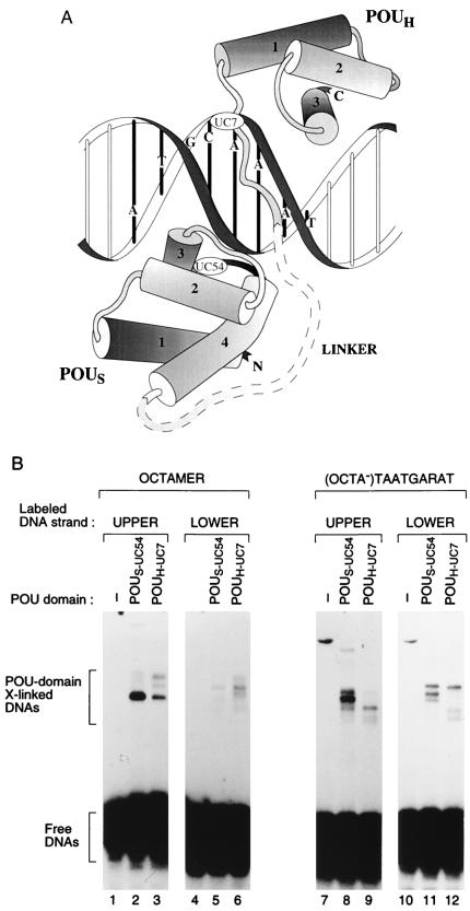
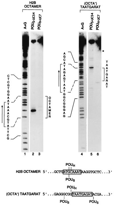
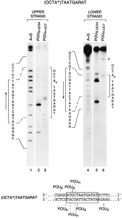
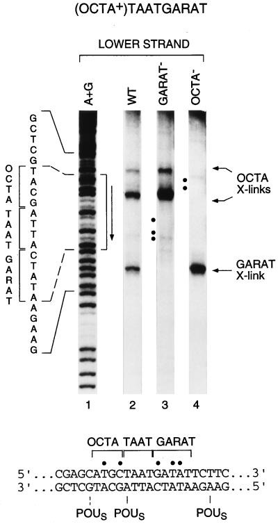
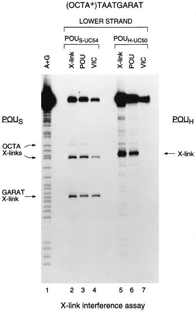
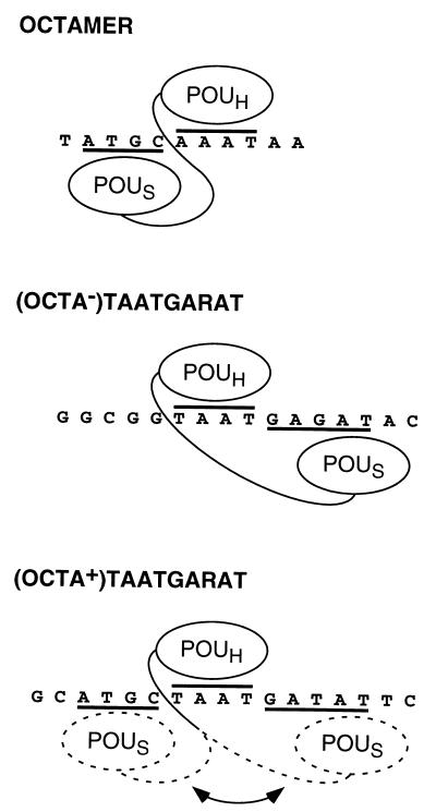
The Oct-1 POU domain binds diverse DNA-sequence elements and forms a higher-order regulatory complex with the herpes simplex virus coregulator VP16. The POU domain contains two separate DNA-binding domains joined by a flexible linker. By protein-DNA photocrosslinking we show that the relative positioning of the two POU DNA-binding domains on DNA varies depending on the nature of the DNA target. On a single VP16-responsive element, the POU domain adopts multiple conformations. To determine the structure of the Oct-1 POU domain in a multiprotein complex with VP16, we allowed VP16 to interact with previously crosslinked POU-domain-DNA complexes and found that VP16 can associate with multiple POU-domain conformations. These results reveal the dynamic potential of a DNA-binding domain in directing transcriptional regulatory complex formation.


Figures

Figure 1
Figure 1
Unique cysteines in the POUS and POUH domains effective for protein–DNA crosslinking. (A) Representation (adapted from refs. and 4) of the Oct-1 POU domain–octamer site crystal structure with the positions of the unique cysteines used for crosslinking analysis indicated (UC54 in the POUS domain and UC7 in the POUH domain; the precise location of UC54 is hidden behind helix 2 on the turn between helices 3 and 4). (B) SDS/PAGE analysis of crosslinked protein–DNA complexes after UV irradiation of POU domain-binding reactions containing no protein (lanes 1, 4, 7, and 10), POUS-UC54 protein (lanes 2, 5, 8, and 11), or POUH-UC7 protein (lanes 3, 6, 9, and 12). Shown are crosslinked complexes formed on the upper and lower strands of the histone H2B octamer and the (OCTA)TAATGARAT sites, as indicated.
Figure 2
Figure 2
The POUS domain is located on opposite sides of the POUH domain on octamer and (OCTA)TAATGARAT sites. Shown are the cleavage products generated after crosslinking of POUS-UC54 and POUH-UC7 proteins to the upper strands of the octamer (lanes 2 and 3) and the (OCTA)TAATGARAT (lanes 5 and 6) sites, as indicated. Lanes 1 and 4 (A+G), purine-cleavage pattern of the probe indicated. In lanes 2, 3, 5, and 6, uppermost bands represent uncleaved DNA. Brackets mark the limits of the two sequences on the autoradiographs, and arrows indicate the directionality of each consensus motif. The two binding sites are cloned into the same plasmid in opposite orientations. ∗, Cleavage product mapping to flanking polylinker sequence, probably resulting from non-octamer-site binding. (Lower) Summary of positions of crosslink-induced cleavage. Dashed line indicates lower-yield cleavage product.
Figure 3
Figure 3
The POU domain adopts multiple conformations on an (OCTA+)TAATGARAT site. Shown are the cleavage products generated from POU domain–DNA crosslinking to both strands of the (OCTA+)TAATGARAT site. Lanes 2 and 3, cleavage pattern generated on the upper strand. The POUH doublet may be due to an incomplete cleavage reaction. Lanes 5 and 6, cleavage pattern generated on the lower strand. Lanes 1 and 4 (A+G), purine-cleavage pattern of the strand indicated. In lanes 5 and 6, the uppermost bands represent uncleaved DNA. Brackets, arrows, and asterisk are as in Fig. 2. (Lower) Summary of positions of cleavage. Dashed lines indicate lower-yield cleavage products.
Figure 4
Figure 4
The POUS domain binds to the wild-type half-site in mutant forms of the (OCTA+)TAATGARAT site. Shown are the cleavage products generated from crosslinking of the POUS domain to wild-type (lane 2) and mutant (lanes 3 and 4) (OCTA+)TAATGARAT sites. Lane 1 (A+G), purine-cleavage pattern of the wild-type probe; lane 3, (OCTA+)TAATGARAT site with the mutated 3′ GARAT sequence (underlined), … ATTAGCTTC… (mutations shown in bold); lane 4, (OCTA+)TAATGARAT site with the mutated 5′ OCTA sequence (underlined), … GCAAGGTA… (mutations shown in bold). Brackets and vertical arrows are as in Fig. 2. (Lower) Sequence with sites of mutations marked by dots and crosslinking sites indicated. Dashed line indicates lower-yield cleavage product.
Figure 5
Figure 5
VP16 associates with multiple conformations of the Oct-1 POU domain. Shown are the results of a crosslinking interference analysis of two unique-cysteine derivatives of the POU domain, POUS-UC54 (lanes 2–4) and POUH-UC50 (lanes 5–7), crosslinked to the lower strand of the (OCTA+)TAATGARAT site. POUS crosslinks over the 5′ ATGC (OCTA X-links) or 3′ GARAT (GARAT X-link) sequences and POUH crosslinks (X-link) are indicated. In lanes 2–7, the uppermost bands represent uncleaved DNA. X-link (above lanes), standard crosslinking analysis of the POU domain alone to the site; POU, crosslinking pattern of the POU-domain complex after electrophoretic mobility retardation; VIC, crosslinking interference pattern of the VP16-induced complex after electrophoretic mobility retardation.
Figure 6
Figure 6
Model for binding of the Oct-1 POU domain to three DNA targets, the octamer and the (OCTA) and (OCTA+) TAATGARAT sites, highlighting conformational flexibility in Oct-1 POU DNA-binding domain arrangement.

Similar articles

Cited by

References

    1. Herr W, Sturm R A, Clerc R G, Corcoran L M, Baltimore D, Sharp P A, Ingraham H A, Rosenfeld M G, Finney M, Ruvkun G, Horvitz H R. Genes Dev. 1988;2:1513–1516. - PubMed
    1. Klemm J D, Rould M A, Aurora R, Herr W, Pabo C O. Cell. 1994;77:21–32. - PubMed
    1. Jacobson E M, Li P, Leon-del-Rio A, Rosenfeld M G, Aggarwal A K. Genes Dev. 1997;11:198–212. - PubMed
    1. Herr W, Cleary M A. Genes Dev. 1995;9:1679–1693. - PubMed
    1. Li P, He X, Gerrero M R, Mok M, Aggarwal A, Rosenfeld M G. Genes Dev. 1993;7:2483–2496. - PubMed

Publication types

MeSH terms

LinkOut - more resources