. 1997 Aug 5;94(16):8691-6.
doi: 10.1073/pnas.94.16.8691.

A family of fibrinogen-related proteins that precipitates parasite-derived molecules is produced by an invertebrate after infection

Affiliations

A family of fibrinogen-related proteins that precipitates parasite-derived molecules is produced by an invertebrate after infection

C M Adema et al. Proc Natl Acad Sci U S A. .

Abstract

After infection with the digenetic trematode Echinostoma paraensei, hemolymph of the snail Biomphalaria glabrata contains lectins comprised of 65-kDa subunits that precipitate polypeptides secreted by E. paraensei intramolluscan larvae. Comparable activity is lacking in hemolymph of uninfected snails. Three different cDNAs with sequence similarities to peptides derived from the 65-kDa lectins were obtained and unexpectedly found to encode fibrinogen-related proteins (FREPs). These FREPs also contained regions with sequence similarity to Ig superfamily members. B. glabrata has at least five FREP genes, three of which are expressed at increased levels after infection. Elucidation of components of the defense system of B. glabrata is relevant because this snail is an intermediate host for Schistosoma mansoni, the most widely distributed causative agent of human schistosomiasis. These results are novel in suggesting a role for invertebrate FREPs in recognition of parasite-derived molecules and also provide a model for investigating the diversity of molecules functioning in nonself-recognition in an invertebrate.


Figures

Figure 1
Figure 1
(A) SDS/PAGE gradient (5–20%) gel of precipitates formed 1.5 h after mixing 0.1, 1.0, 2.5, 5, 10, 25, or 50 μl of SEP with different 25-μl aliquots of a common plasma pool. Shown beneath the gel is a graph of protein content (micrograms) of precipitates from each lane. The results are explained in the text. Note the presence of a 49-kDa band derived from SEP in lanes receiving higher doses of SEP. Mr markers (kDa) are indicated. (B) SDS/PAGE gradient (5–20%) gel showing that, after exposure to SEP-free medium, both ppt 1 and ppt 2 remained in an insoluble form in a pellet (lane 1); they were not solubilized, and thus were absent from the supernatant (lane 2). In contrast, after exposure to SEP, components of ppt 1 and ppt 2 were solubilized and thus were not retained in the pellet (lane 3) but appeared in the supernatant (lane 4). Polypeptides smaller than 65 kDa are from SEP. Mr markers (kDa) are indicated.
Figure 2
Figure 2
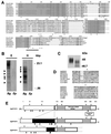
(A) Alignment of gastropod FREP domains with HsaFibB: Homo sapiens fibrinogen B, M64983; HsaP35: H. sapiens plasma lectin P35, D49353; PpaFREP: FREP Parastichopus parvimensis (sea cucumber), M31326; and LflSASL: Limax flavus (slug) sialic acid-specific lectin, A48505. Note the fibrinogen characteristics of deduced amino acid sequences of the B. glabrata FREPs BgMFREP1–4 and the Bulinus truncatus FREP (BtrFREP1) provided as an example of the other gastropod FREPs obtained. Identical amino acid residues are bolded. Stars indicate amino acids conserved among fibrinogen-related sequences (13). In all available snail sequences, the tryptophan (W) in position 84 has undergone a conserved replacement with a tyrosine (Y). Similarly, a Y (position 212) was replaced by a W. The spacings between residues 68 and 80 as well as 106 and 108 are snail-specific. The two shaded boxes indicate areas that are homologous to the calcium-binding sites that are the likely carbohydrate-binding domains of the human serum lectin P35 (15). The Prosite motif (PS00514) for fibrinogen is underlined: BgMFREP3 has one additional residue at position 203. Residues have been renumbered to allow comparison of the FREP domains. (B) Genomic DNA from one B. glabrata snail (Bg) and from E. paraensei sporocysts (Ep) was digested with EcoRI (E) or HaeIII (H) and probed at high stringency with radiolabeled BgMFREP1. The probe hybridized with at least five B. glabrata DNA fragments (arrowheads). No signal was detected from E. paraensei DNA. (C) Reduction in Mr of the 65-kDa band (Left) to 46.7-kDa band (Right) after 24 h of deglycosylation with N-glycosidase F. (D) Partial sequences from the 5′ nonfibrinogen-related portion of B. glabrata FREPs have homologies to parts of Ig superfamily molecules. In particular, two cysteines that are situated 80 residues sequences apart are both flanked by residues that align with residues around intra-chain, Ig-type, loop-forming cysteines. Alignments are shown for the cysteine residues (indicated by stars) present at both positions 31 (Upper) and 111 (Lower) of the complete BgMFREP2 sequence. Relative to the BgMFREP2 sequence, identical residues are bolded, and conserved replacements are italicized. Abbreviations and GenBank accession numbers of the Ig superfamily sequences used for the alignments in this figure: CelCAM: Caenorhabditis elegans, similar to C2-type domains of N-CAM proteins, U29082; GgaCAM-GRASP: Gallus gallus (chicken) cellular adhesion molecule GRASP, JH0464; HsaIgEVR: Homo sapiens IgE variable region, Z26839; HsaIgKCVR: Homo sapiens Ig κ chain variable region, L03684; HsaIgLCVR: Homo sapiens Ig light chain variable region, X13084; HsaTCRβVR: Homo sapiens T cell receptor β variable region, D13086; RnoTCRβVR Rattus norvegicus T cell receptor β variable region, B30563; MamTCRαVR: Macaca mulatta T cell receptor α variable region, U11793; MmuIgHCVR: Mus musculus Ig heavy chain variable region, S36380; LstMDM, Lymnaea stagnalis (pond snail), molluscan defense molecule, U58769; and OcuTCRβVR: Oryctolagus cuniculus (rabbit), T cell receptor β variable region, d17420. (E) Schematic alignment of (partial) BgMFREPs shows that FREP domains are located at the 3′ end of each sequence, immediately upstream from a stop codon. Gray boxes indicate the positions of two putative calcium-dependent carbohydrate-binding sequences. At the 5′ end, BgMFREPs show homologies to each other, but similarities with any other known genes are minimal. The length of the interceding sequence between these two regions varied between BgMFREPs (black boxes); BgMFREP2 is the shortest B. glabrata FREP obtained. Arrowheads indicate N-linked glycosylation sites (PS00001, Prosite). The position of the Prosite motif is also indicated.
Figure 3
Figure 3
(A) Northern blot of total RNA from an individual infected (I) or uninfected (U) control snail. Radiolabeled BgMFREP1 hybridized with at least five transcripts in RNA from the infected snail (arrow heads). Fewer and lighter transcripts were detected in RNA from the uninfected snail. The probe BgMFREP2 hybridized strongly with a 1450-nt transcript. No BgMFREP2-specific signal was detected from the uninfected snail. (B) RNA samples from infected (I) and uninfected (U) snails (two examples of each shown) served as template for RT-PCR reactions using BgMFREP1–3-specific sense and antisense primers. For each primer pair, the graph depicts the number of positive signals from 10 infected and 10 uninfected snails. BgMEST4 is a housekeeping gene control. (C) Slot blot Northern blotting analyses of total RNA from individual infected (I) and uninfected (U) snails were probed with either BgMFREP1+2 or BgMEST4, a housekeeping gene. Densitometric intensity of BgMFREP-specific signal [expressed as arbitrary units (AU)] relative to the BgMEST4-dependent signal was calculated for six infected and five control snails. The mean signal strength was significantly higher in infected snails (Student’s t test, P = 0.0025).

References

    1. Lemaitre B, Nicolas E, Michaut L, Reichart J-M, Hoffmann J A. Cell. 1996;86:973–983. - PubMed
    1. Richards E H, Renwrantz L R. J Comp Physiol. 1991;161:43–54.
    1. Van der Knaap W P W, Loker E S. Parasitol Today. 1990;6:175–182. - PubMed
    1. Adema C M, Hertel L A, Loker E S. In: Parasites and Pathogens: Effects on Host Hormones and Behavior. Beckage N, editor. New York: Chapman & Hall; 1997. pp. 76–98.
    1. Hertel L A, Stricker S A, Monroy F P, Wilson W D, Loker E S. J Invert Pathol. 1994;64:52–61. - PubMed

Publication types