. 1997 Aug 5;94(16):8756-60.
doi: 10.1073/pnas.94.16.8756.

Resistance to apoptosis caused by PIG-A gene mutations in paroxysmal nocturnal hemoglobinuria

Affiliations

Resistance to apoptosis caused by PIG-A gene mutations in paroxysmal nocturnal hemoglobinuria

R A Brodsky et al. Proc Natl Acad Sci U S A. .

Abstract

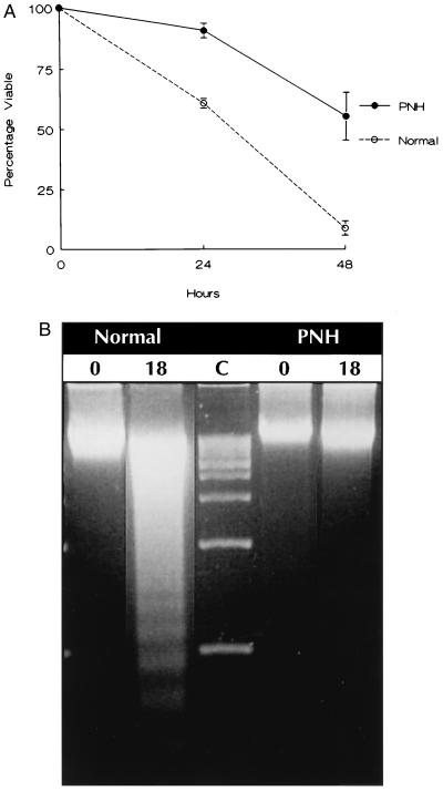
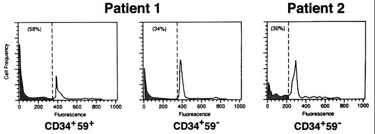
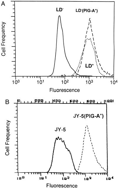
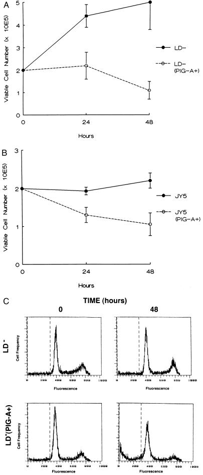
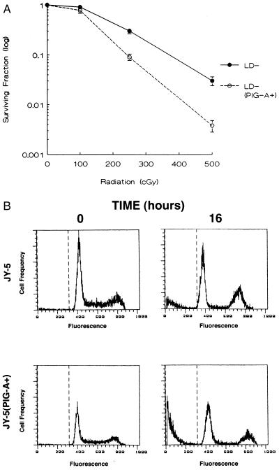
Paroxysmal nocturnal hemoglobinuria (PNH) is a clonal hematopoietic stem cell disorder resulting from mutations in an X-linked gene, PIG-A, that encodes an enzyme required for the first step in the biosynthesis of glycosylphosphatidylinositol (GPI) anchors. PIG-A mutations result in absent or decreased cell surface expression of all GPI-anchored proteins. Although many of the clinical manifestations (e.g., hemolytic anemia) of the disease can be explained by a deficiency of GPI-anchored complement regulatory proteins such as CD59 and CD55, it is unclear why the PNH clone dominates hematopoiesis and why it is prone to evolve into acute leukemia. We found that PIG-A mutations confer a survival advantage by making cells relatively resistant to apoptotic death. When placed in serum-free medium, granulocytes and affected CD34(+) (CD59(-)) cells from PNH patients survived longer than their normal counterparts. PNH cells were also relatively resistant to apoptosis induced by ionizing irradiation. Replacement of the normal PIG-A gene in PNH cell lines reversed the cellular resistance to apoptosis. Inhibited apoptosis resulting from PIG-A mutations appears to be the principle mechanism by which PNH cells maintain a growth advantage over normal progenitors and could play a role in the propensity of this disease to transform into more aggressive hematologic disorders. These data also suggest that GPI anchors are important in regulating apoptosis.


Figures

Figure 1
Figure 1
(A) Survival of PNH and normal peripheral blood granulocytes in serum-free medium. Cell viability was determined by trypan blue dye exclusion. Each data point indicating percent survival represents the ratio of the absolute viable cell number at the time of assay and the number of viable cells at the start of each experiment. The data are the mean ± SEM of four patients with PNH and five normals. (B) Gel electrophoresis for detection of oligonucleosomal DNA fragments at time 0 and 18 hr after culture of normal and PNH granulocytes in serum-free medium. Lane C depicts 1-kb DNA size markers.
Figure 2
Figure 2
Flow cytometric analysis for apoptosis of CD34+ cells from two PNH patients. Unaffected (CD59+) and affected (CD59) cells from patient 1, and affected cells from patient 2. The number in parenthesis indicates the percentage of subdiploid DNA (shaded).
Figure 3
Figure 3
Flow cytometric analysis for CD59 expression. (A) The GPI anchor-deficient, LD (solid line), and the GPI anchor-replete, LD+ (dotted line), Epstein–Barr virus-transformed B lymphoblastoid cell lines were derived from a patient with PNH. LD (PIG-A+) (dashed line) was established after stable transfection of the PIG-A cDNA into the LD cell line. (B) The GPI anchor-deficient JY-5 cells (solid line) and JY-5 (PIG-A+) cells (dashed line).
Figure 4
Figure 4
Survival of PNH cells lines in serum-free medium. (A) LD and LD (PIG-A+). (B) JY-5 and JY-5 (PIG-A+). Cell viability was determined by trypan blue exclusion after incubation in serum-free medium for 48 hr. The data are the mean of ± SEM of three separate experiments. (C) Flow cytometric analysis for apoptosis of LD and LD (PIG-A+) cells after 48 hr in serum-free medium.
Figure 5
Figure 5
Relative sensitivity of PNH cell lines to irradiation. (A) The surviving fraction from LD and LD (PIG-A+) cells represents the number of clonogenic colonies after treatment compared with the corresponding untreated controls. Each data point represents the mean ± SEM of three separate experiments. (B) Since JY-5 cells were unable to be cloned in semi-solid medium, flow cytometric analysis was used to assess apoptosis after 500 cGy of ionizing radiation. Propidium iodide staining of the JY-5 cells for DNA content revealed 10.5% ± 2.3 subdiploid DNA 16 hr after radiation. In contrast, JY-5 (PIG-A+) cells revealed 31.3% ± 2.9 subdiploid DNA after identical conditions. A representative example from one of the three experiments is shown.

References

    1. Hillmen P, Lewis S M, Bessler M, Luzzatto L, Dacie J V. N Engl J Med. 1995;333:1253–1258. - PubMed
    1. Rotoli B, Luzzatto L. Bailliere’s Clin Haematol. 1989;2:113–138. - PubMed
    1. Takeda J, Miyata T, Kawagoe K, Iida Y, Endo Y, Fujita T, Takahashi M, Kitani T, Kinoshita T. Cell. 1993;73:703–711. - PubMed
    1. Miyata T, Yamada N, Iida Y, Nishimura J, Takeda J, Kitani T, Kinoshita T. N Engl J Med. 1994;330:249–255. - PubMed
    1. Bessler M, Mason P J, Hillmen P, Miyata T, Yamada N, Takeda J, Luzzatto L, Kinoshita T. EMBO J. 1994;13:110–117. - PMC - PubMed

Publication types

MeSH terms

Substances

LinkOut - more resources