. 1997 Aug 5;94(16):8807-11.
doi: 10.1073/pnas.94.16.8807.

Loss of virulence in Leishmania donovani deficient in an amastigote-specific protein, A2

Affiliations

Loss of virulence in Leishmania donovani deficient in an amastigote-specific protein, A2

W W Zhang et al. Proc Natl Acad Sci U S A. .

Abstract

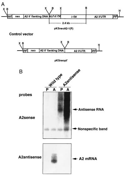
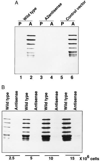
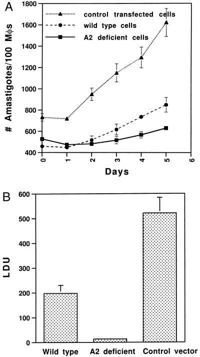
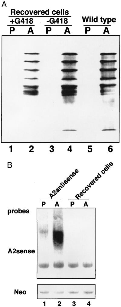
Leishmania donovani is the etiologic agent of fatal visceral leishmaniasis in man. During their life cycle, Leishmania exist as flagellated promastigotes within the sandfly vector and as nonflagellated amastigotes in the macrophage phagolysosomal compartment of the mammalian host. The transformation from promastigotes to amastigotes is a critical step for the establishment of infection, and the molecular basis for this transformation is poorly understood. To define the molecular basis for amastigote survival in the mammalian host, we previously identified an amastigote stage-specific gene family termed "A2." In the present study, we have inhibited the expression of A2 mRNA and A2 protein in amastigotes using antisense RNA and show that the resulting A2-deficient amastigotes are severely compromised with respect to virulence in mice. Amastigotes that did survive in the mice had restored A2 protein expression. These data demonstrate that A2 protein is required for L. donovani survival in a mammalian host, and this represents the first identified amastigote-specific virulence factor identified in Leishmania. This study also reveals that it is possible to study gene function in Leishmania through the expression of antisense RNA.


Figures

Figure 1
Figure 1
(A) Structures of the A2 antisense RNA expressing vector (pKSneoA2–1 R) construct and the control vector. The reversed 2.4-kb sequence containing 1.6 kb of A2 encoding sequence and part of the 3′ UTR was flanked by a 1.6-kb A2 5′ noncoding upstream sequence and 1.7 kb of A2 3′ UTR. Pyt is a 92-bp synthetic pyrimidine tract that provides the trans-splicing site for the neor gene and insures the correct polyadenylylation of antisense transcripts at the end of A2 3′ UTR. The 2.4-kb BamHI fragment containing A2–1 encoding sequence was removed in the control vector (pKSneopt). Restriction enzyme sites are indicated: X, XbaI; B, BamHI; H, XhoI. (B) Northern blot analysis of RNA isolated from wild-type and pKSneoA2–1 R plasmid-transfected Leishmania cells. Total RNAs (10 μg per lane) isolated from promastigotes (P) or amastigotes (A) from wild-type or transfected cells were subjected to Northern blot analysis with a sense oligonucleotides specific for A2 antisense RNAs (Upper) or an antisense oligonucleotides specific for native A2 mRNAs (Lower). Note: A2 antisense RNAs were detected only in cells transfected with the A2 antisense-expressing plasmid and almost no native A2 mRNA was detected in the amastigotes containing the A2 antisense RNA. Higher levels of A2 antisense RNAs were present in amastigotes than in promastigotes because these transcripts contained the A2 3′ UTR.
Figure 2
Figure 2
(A) Western blot analysis of the A2 amastigote-specific proteins in wild-type and transfected Leishmania cells; P, promastigotes; A, amastigotes. Equal amounts of total protein extracted from 5 × 10⁶ cells were subjected to Western blot analysis with an anti-A2 mAb. Note: There is a dramatic reduction in the amount of A2 protein in the amastigotes containing the A2 antisense RNA (lane 4) whereas the wild-type amastigotes (lane 2) or amastigotes containing the control vector (lane 6) contain relatively high levels of A2. (B) A2 protein levels were compared in total proteins extracted from increasing numbers of wild-type amastigotes and amastigotes expressing the A2 antisense RNA as indicated.
Figure 3
Figure 3
(A) The infection and growth rates of wild-type and transfected Leishmania amastigotes in macrophages in vitro. BALB/c mouse bone marrow-derived macrophages were infected at an amastigote-to-cell ratio of 20:1 for 24 h. Noningested amastigotes were removed by centrifugation. The infected cells were incubated at 37°C. Amastigote infection level and growth rate in macrophages were evaluated daily by cytospin and Giemsa staining. The data are the mean ± SEM (bar) of three independent experiments. (B) Liver parasite burdens of mice infected with wild-type and transfected Leishmania cells as indicated. Female BALB/c mice were injected with amastigotes via the tail vein (1.5 × 10⁸ amastigotes/mouse, three mice per group). Four weeks after infection, mice were examined for hepatic parasite burdens by counting amastigotes from liver impression. Liver parasite burdens, expressed as Leishman–Donovan units (LDU), were calculated as follows: number of amastigotes per 1000 cell nuclei × liver weight (g). The means per group are shown, with the error bars representing SE. These results are from four separate experiments, each which gave very reproducible results. Similar results were obtained when mice were directly infected with wild-type and transfected promastigotes.
Figure 4
Figure 4
(A) A2 protein levels are restored in amastigotes isolated from liver and spleen 4 weeks after mice were infected with A2-deficient amastigotes. The amastigotes isolated from infected mice were cultured in vitro in promastigote or amastigote culture medium with or without 20 μg/ml G418. The cells were collected after they reached a density of 3 × 10⁷/ml and were subjected to Western Blot analysis with an anti-A2 mAb. P, promastigotes; A, amastigotes. For comparison, the levels of A2 proteins in wild-type promastigotes and amastigotes are also shown. (B) A2 antisense RNA is lost, but neo RNA is retained in amastigotes isolated from liver and spleen 4 weeks after mice were infected with amastigotes expressing both A2 antisense RNA and neo RNA. Cells were prepared as described above, and Northern blot analysis was performed using probes specific for A2 antisense RNA and neo RNA. P, promastigotes; A, amastigotes.

Similar articles

Cited by

References

    1. Molyneux D, Killick-Kendrick R. In: The Leishmaniases in Biology and Medicine. Peters W, Killick-Kendrick R, editors. Vol. 1. London: Academic; 1987. pp. 121–176.
    1. Schlein Y. Parasitol Today. 1993;9:255–258. - PubMed
    1. Charest H, Matlashewski G. Mol Cell Biol. 1994;14:2975–2984. - PMC - PubMed
    1. Charest H, Zhang W-W, Matlashewski G. J Biol Chem. 1996;271:17081–17090. - PubMed
    1. Zhang W-W, Charest H, Ghedin E, Matlashewski G. Mol Biochem Parasitol. 1996;78:79–90. - PubMed

Publication types

LinkOut - more resources