. 1997 Aug 5;94(16):8818-23.
doi: 10.1073/pnas.94.16.8818.

Intrastriatal injection of an adenoviral vector expressing glial-cell-line-derived neurotrophic factor prevents dopaminergic neuron degeneration and behavioral impairment in a rat model of Parkinson disease

Affiliations

Intrastriatal injection of an adenoviral vector expressing glial-cell-line-derived neurotrophic factor prevents dopaminergic neuron degeneration and behavioral impairment in a rat model of Parkinson disease

A Bilang-Bleuel et al. Proc Natl Acad Sci U S A. .

Abstract

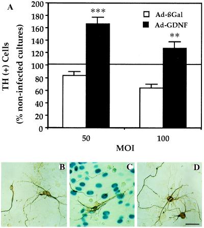
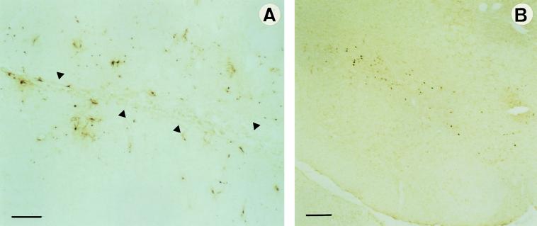
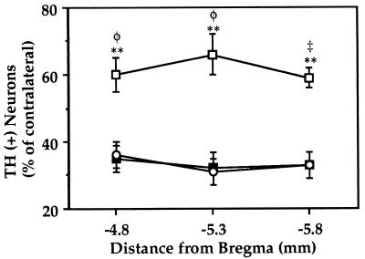
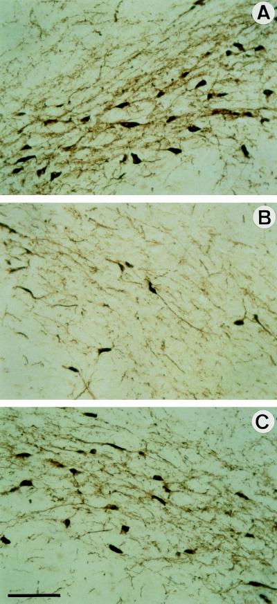
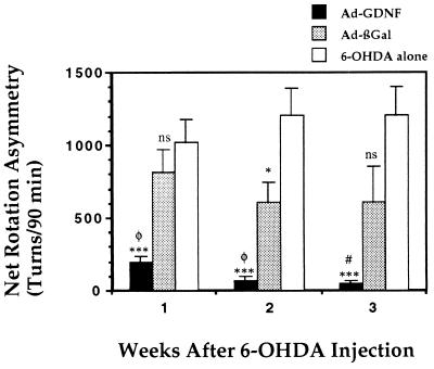
Glial-cell-line-derived neurotrophic factor (GDNF) is a potent neurotrophic factor for adult nigral dopamine neurons in vivo. GDNF has both protective and restorative effects on the nigro-striatal dopaminergic (DA) system in animal models of Parkinson disease. Appropriate administration of this factor is essential for the success of its clinical application. Since it cannot cross the blood-brain barrier, a gene transfer method may be appropriate for delivery of the trophic factor to DA cells. We have constructed a recombinant adenovirus (Ad) encoding GDNF and injected it into rat striatum to make use of its ability to infect neurons and to be retrogradely transported by DA neurons. Ad-GDNF was found to drive production of large amounts of GDNF, as quantified by ELISA. The GDNF produced after gene transfer was biologically active: it increased the survival and differentiation of DA neurons in vitro. To test the efficacy of the Ad-mediated GDNF gene transfer in vivo, we used a progressive lesion model of Parkinson disease. Rats received injections unilaterally into their striatum first of Ad and then 6 days later of 6-hydroxydopamine. We found that mesencephalic nigral dopamine neurons of animals treated with the Ad-GDNF were protected, whereas those of animals treated with the Ad-beta-galactosidase were not. This protection was associated with a difference in motor function: amphetamine-induced turning was much lower in animals that received the Ad-GDNF than in the animals that received Ad-beta-galactosidase. This finding may have implications for the development of a treatment for Parkinson disease based on the use of neurotrophic factors.


Figures

Figure 1
Figure 1
Increased survival of DA neurons in cultures infected with Ad-GDNF. (A) DA-cell counts by TH immunocytochemistry. Embryonic mesencephalic cells (100,000 cells per cm2) were plated on a layer of primary astroglial cells (100,000 cells per cm2) previously infected with Ad-GDNF or Ad-βGal. DA cells were identified with TH immunocytochemistry and counted. The values represent the percentages of total DA cells counted in noninfected cultures (means ± SEM of three determinations). In the control noninfected cultures, there were 982 ± 47 DA cells per well. ∗∗, P < 0.01; ∗∗∗, P < 0.001 versus Ad-βGal. (B–D) Differentiation of DA neurons visualized by TH immunocytochemistry. Photomicrographs illustrating the morphology of DA cells plated on a layer of primary astroglial cells previously or not infected. (B) Noninfected astrocytes, (C) astrocytes infected with Ad-ßGal (50 pfu per cell), (D) with Ad-GDNF (50 pfu per cell). βGal expression is visualized enzymatically with 5-bromo-4-chloro-3-indolyl β-d-galactoside as a substrate giving rise to a blue color. Note that the differentiation of DA neurons is much more advanced in cultures producing the transgenic GDNF than in control or Ad-βGal-infected cultures. (Bar = 100 μm.)
Figure 2
Figure 2
Analysis of adenoviral transgene expression using βGal immunohistochemistry. Pictures of 14-μm-thick coronal sections through the caudate putamen (A) and SN (B) showing βGal-expressing cells 4 weeks after intrastriatal injection of Ad-βGal. Anti-E. coli βGal antibodies were used to distinguish the transgenic from the endogenous βGal activity. In the striatum, βGal+ cells are found along the needle tract (indicated by arrows in A) and up to 2 mm from the site of injection. Numerous infected cells can be observed in the SN compacta (B) after retrograde transport of viral particles delivered in the caudate putamen. (Bar = 200 μm.)
Figure 3
Figure 3
Survival of DA neurons in the SN of 6-OHDA lesioned rats. The animals received intrastriatal Ad injections followed by 6-OHDA 6 days later. Three weeks after 6-OHDA injection, animals were sacrificed for TH immunohistochemistry. The number of TH+ cell bodies present in the SN at the coordinates AP −4.8, −5.3, and −5.8 mm from bregma (three or four sections per region of each animal) was determined. The values reported are means ± SEM for 6–11 rats per group and are expressed as percentages of TH+ cell counts in the contralateral nonlesioned SN. The survival of DA neurons is significantly higher in animals injected with Ad-GDNF (□) than with Ad-βGal (▪) or than in animals that received 6-OHDA alone (○). ∗∗, P < 0.01 versus 6-OHDA alone; φ, P < 0.01 and , P < 0.001 versus Ad-βGal.
Figure 4
Figure 4
Histological analysis of SN DA neurons of treated rats. Representative pictures of 14-μm-thick coronal sections through the SN processed for TH immunohistochemistry are shown. (A) Section contralateral to the lesion. (B and C) Sections ipsilateral to the lesion of animals injected with Ad-βGal (B) or Ad-GDNF (C). The aspect of the ipsilateral SN from rats that received only 6-OHDA is comparable to those of rats that received 6-OHDA and Ad-βGal (see B). (Bar = 100 μm.) The number of TH+ cell bodies and density of TH-stained fibers were both higher in rats that received Ad-GDNF than Ad-βGal (C versus B).
Figure 5
Figure 5
Effect of Ad-GDNF on amphetamine-induced rotational behavior in 6-OHDA-lesioned rats. Ad-GDNF (n = 7) or Ad-βGal (n = 8) were delivered into the left striatum of animals by stereotaxic injection. Six days thereafter, 20 μg of 6-OHDA⋅HCl was injected into the left striatum of all two groups of animal. A third group of animal received no preinjection before 6-OHDA lesion (6-OHDA only, n = 10). The ability of the different treatments to counteract the neurotoxin action was assessed by following asymmetric rotational behavior induced by amphetamine administration 1, 2, and 3 weeks after 6-OHDA injection. The values reported are the means ± SEM (bars) of net ipsilateral turns over 90 min (turns contralateral to the lesion subtracted). ∗, P < 0.05; ∗∗∗, P < 0.001; and ns, not significant versus 6-OHDA alone. #, P < 0.05 and φ, P < 0.01 versus Ad-βGal.

References

    1. Lin L F, Doherty D H, Lile J D, Bektesh S, Collins F. Science. 1993;260:1130–1132. - PubMed
    1. Hou J G, Lin L F, Mytilineou C. J Neurochem. 1996;66:74–82. - PubMed
    1. Sauer H, Rosenblad C, Björklund A. Proc Natl Acad Sci USA. 1995;92:8935–8939. - PMC - PubMed
    1. Beck K D, Valverde J, Alexi T, Poulsen K, Moffat B, Vandlen R A, Rosenthal A, Hefti F. Nature (London) 1995;373:339–341. - PubMed
    1. Tomac A, Widenfalk J, Lin L-F, Kohno T, Ebendal T, Hoffer B J, Olson L. Proc Natl Acad Sci USA. 1995;92:8274–8278. - PMC - PubMed

Publication types

MeSH terms

Substances