. 1997 Aug 11;138(3):485-94.
doi: 10.1083/jcb.138.3.485.

Mutational analysis of Mdm1p function in nuclear and mitochondrial inheritance

Affiliations

Mutational analysis of Mdm1p function in nuclear and mitochondrial inheritance

H A Fisk et al. J Cell Biol. .

Abstract

Nuclear and mitochondrial transmission to daughter buds of Saccharomyces cerevisiae depends on Mdm1p, an intermediate filament-like protein localized to numerous punctate structures distributed throughout the yeast cell cytoplasm. These structures disappear and organelle inheritance is disrupted when mdm1 mutant cells are incubated at the restrictive temperature. To characterize further the function of Mdm1p, new mutant mdm1 alleles that confer temperature-sensitive growth and defects in organelle inheritance but produce stable Mdm1p structures were isolated. Microscopic analysis of the new mdm1 mutants revealed three phenotypic classes: Class I mutants showed defects in both mitochondrial and nuclear transmission; Class II alleles displayed defective mitochondrial inheritance but had no effect on nuclear movement; and Class III mutants showed aberrant nuclear inheritance but normal mitochondrial distribution. Class I and II mutants also exhibited altered mitochondrial morphology, possessing primarily small, round mitochondria instead of the extended tubular structures found in wild-type cells. Mutant mdm1 alleles affecting nuclear transmission were of two types: Class Ia and IIIa mutants were deficient for nuclear movement into daughter buds, while Class Ib and IIIb mutants displayed a complete transfer of all nuclear DNA into buds. The mutations defining all three allelic classes mapped to two distinct domains within the Mdm1p protein. Genetic crosses of yeast strains containing different mdm1 alleles revealed complex genetic interactions including intragenic suppression, synthetic phenotypes, and intragenic complementation. These results support a model of Mdm1p function in which a network comprised of multimeric assemblies of the protein mediates two distinct cellular processes.


Figures
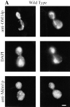
Figure 1
Figure 1
New mdm1 alleles define three phenotypic classes. MYY290 (MDM1) (A), MYY720 (mdm1-251) (B), MYY721 (mdm1-252) (C), and MYY702 (mdm1-200) (D) cells were grown on YPD medium at 23°C, incubated at 37°C for 4 h, fixed with formaldehyde, and processed for indirect immunofluorescence microscopy. Mitochondria were detected with a mouse monoclonal antibody against OM14 (a mitochondrial outer-membrane protein) followed by fluorescein-conjugated goat anti–mouse IgG. Nuclear and mitochondrial DNAs were visualized by DAPI staining. Mdm1p structures were detected using affinity-purified, anti-Mdm1p antibodies followed by rhodamine-conjugated donkey anti–rabbit IgG. For each strain, two representative cells are shown. Bars, 2 μm.
Figure 1
Figure 1
New mdm1 alleles define three phenotypic classes. MYY290 (MDM1) (A), MYY720 (mdm1-251) (B), MYY721 (mdm1-252) (C), and MYY702 (mdm1-200) (D) cells were grown on YPD medium at 23°C, incubated at 37°C for 4 h, fixed with formaldehyde, and processed for indirect immunofluorescence microscopy. Mitochondria were detected with a mouse monoclonal antibody against OM14 (a mitochondrial outer-membrane protein) followed by fluorescein-conjugated goat anti–mouse IgG. Nuclear and mitochondrial DNAs were visualized by DAPI staining. Mdm1p structures were detected using affinity-purified, anti-Mdm1p antibodies followed by rhodamine-conjugated donkey anti–rabbit IgG. For each strain, two representative cells are shown. Bars, 2 μm.
Figure 2
Figure 2
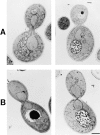
Class II alleles display altered mitochondrial morphology and defects in mitochondrial inheritance. MYY290 (MDM1) (A) and MYY721 (mdm1-252) (B) cells were grown in YPG medium overnight at 23°C, incubated at 37°C in YPG for 6 h, prefixed with glutaraldehyde, fixed in KMnO4, and stained in uranyl acetate. Sections were stained with lead citrate. Two representative cells of each strain are shown. Bar, 1 μm.
Figure 3
Figure 3
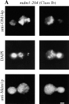
Class Ib-mdm1 and Class IIIb-mdm1 cells display complete transfer of nuclear DNA to daughter buds. MYY709 (mdm1-204) (A) and MYY715 (mdm1-227) (B and C) cells were grown at 23°C, incubated for 4 h at 37°C, and fixed and processed as described for Fig. 1. (A and B) Cells were stained with anti-OM14 followed by fluorescein-conjugated goat anti–mouse IgG, DAPI, and anti-MDM1p followed by rhodamine-conjugated donkey anti–rabbit IgG. (C) Cells were stained with anti-OM14 followed by fluorescein-conjugated goat anti–mouse IgG, DAPI and calcofluor (Calc.; to visualize bud scars), and anti-tubulin followed by rhodamine-conjugated donkey anti–rat IgG. Two representative cells are shown for each strain. Bars, 2 μm.
Figure 3
Figure 3
Class Ib-mdm1 and Class IIIb-mdm1 cells display complete transfer of nuclear DNA to daughter buds. MYY709 (mdm1-204) (A) and MYY715 (mdm1-227) (B and C) cells were grown at 23°C, incubated for 4 h at 37°C, and fixed and processed as described for Fig. 1. (A and B) Cells were stained with anti-OM14 followed by fluorescein-conjugated goat anti–mouse IgG, DAPI, and anti-MDM1p followed by rhodamine-conjugated donkey anti–rabbit IgG. (C) Cells were stained with anti-OM14 followed by fluorescein-conjugated goat anti–mouse IgG, DAPI and calcofluor (Calc.; to visualize bud scars), and anti-tubulin followed by rhodamine-conjugated donkey anti–rat IgG. Two representative cells are shown for each strain. Bars, 2 μm.
Figure 4
Figure 4
Nuclear distribution defects in Class III-mdm1 cells. MYY290 (MDM1), Class IIIa mutants MYY702 (mdm1-200) and MYY710 (mdm1-217), and Class IIIb mutants MYY715 (mdm1-227) and MYY717 (mdm1-228) were synchronized with α-factor at 23°C, released from the cell cycle block at 37°C, fixed with formaldehyde after 0, 2, or 4 h, and stained with DAPI. For each sample, the distribution of phenotypes among 300 cells was determined. Values represent the mean ± standard deviation for triplicate samples. Between 85 and 95% of cells in 0 h samples were unbudded. Inset shows symbols corresponding to each culture, and each phenotype is represented below the x axis. (A) 2 h after release at 37°C. (B) 4 h after release at 37°C.
Figure 5
Figure 5
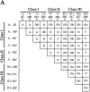
Intragenic interaction among mdm1 alleles. Genetic crosses were performed to obtain all possible pairwise combinations of different mdm1 alleles. Organelle inheritance phenotypes were analyzed as described in Fig. 1. (A) Phenotypic classes of mdm1 diploids. Numbers on the x and y axis indicate mdm1 allele of parental strains. Roman numeral refers to phenotypic class, and lower case “a” or “b” refers to nuclear transmission phenotype, as described in Table II and in the text. “R” or “D” indicates the recessive or dominant character of individual alleles. (B) Intragenic complementation is observed between mdm1-217 (Class IIIa) and mdm1-228 (Class IIIb). Haploids and diploids harboring the mdm1-217 and mdm1-228 mutations were tested for growth on YPD at 37°C. (a–c) mdm1-217; (a) MATa, (b) MATα, and (c) MATa/α; (d–f) mdm1-228; (d) MATa, (e) MATα, and (f) MATa/α; (g) mdm1-217/mdm1-228 (a/α); (h) mdm1-228/mdm1-217.
Figure 5
Figure 5
Intragenic interaction among mdm1 alleles. Genetic crosses were performed to obtain all possible pairwise combinations of different mdm1 alleles. Organelle inheritance phenotypes were analyzed as described in Fig. 1. (A) Phenotypic classes of mdm1 diploids. Numbers on the x and y axis indicate mdm1 allele of parental strains. Roman numeral refers to phenotypic class, and lower case “a” or “b” refers to nuclear transmission phenotype, as described in Table II and in the text. “R” or “D” indicates the recessive or dominant character of individual alleles. (B) Intragenic complementation is observed between mdm1-217 (Class IIIa) and mdm1-228 (Class IIIb). Haploids and diploids harboring the mdm1-217 and mdm1-228 mutations were tested for growth on YPD at 37°C. (a–c) mdm1-217; (a) MATa, (b) MATα, and (c) MATa/α; (d–f) mdm1-228; (d) MATa, (e) MATα, and (f) MATa/α; (g) mdm1-217/mdm1-228 (a/α); (h) mdm1-228/mdm1-217.
Figure 6
Figure 6
Distribution of mutations in Mdm1p.
Figure 7
Figure 7
Summary of mdm1 mutant phenotypes.

Similar articles

Cited by

References

    1. Adams RJ, Pollard TD. Propulsion of organelles isolated from Acanthamoeba along actin filaments by myosin-I. Nature (Lond) 1986;322:754–756. - PubMed
    1. Allan V. Membrane traffic motors. FEBS (Fed Eur Biochem Soc) Lett. 1995;369:101–106. - PubMed
    1. Ball EH, Singer SJ. Mitochondria are associated with microtubules and not with intermediate filaments in cultured fibroblasts. Proc Natl Acad Sci USA. 1982;79:123–126. - PMC - PubMed
    1. Berger KH, Yaffe MP. Mitochondrial distribution and inheritance. Experientia (Basel) 1996;52:1111–1116. - PubMed
    1. Chen LB. Mitochondrial membrane potential in living cells. Annu Rev Cell Biol. 1988;4:155–181. - PubMed

Publication types

MeSH terms

Substances