. 1997 Aug 11;138(3):517-29.
doi: 10.1083/jcb.138.3.517.

A multispecificity syntaxin homologue, Vam3p, essential for autophagic and biosynthetic protein transport to the vacuole

Affiliations

A multispecificity syntaxin homologue, Vam3p, essential for autophagic and biosynthetic protein transport to the vacuole

T Darsow et al. J Cell Biol. .

Abstract

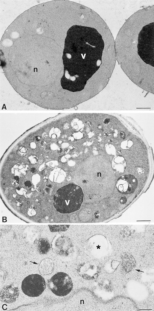
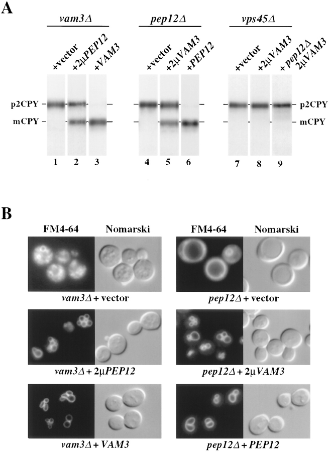
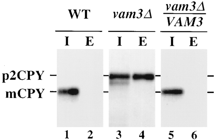
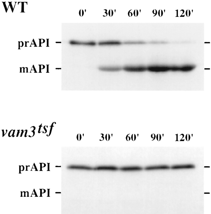
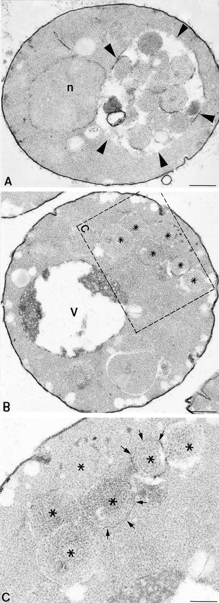
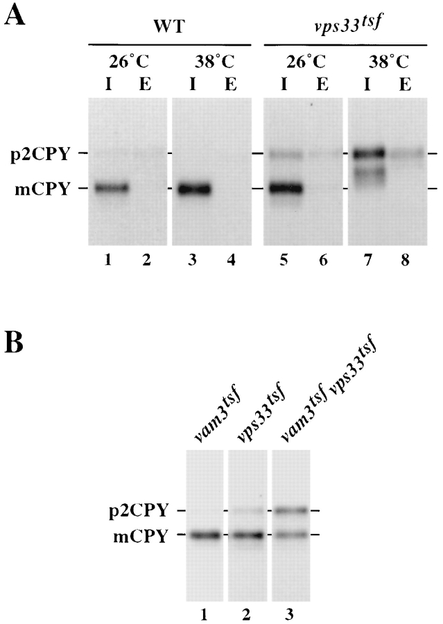
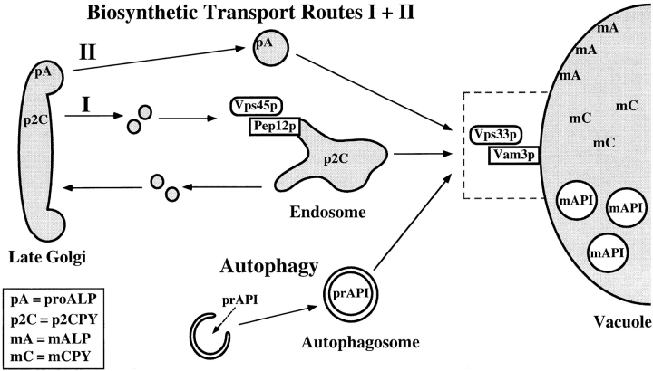
Protein transport in eukaryotic cells requires the selective docking and fusion of transport intermediates with the appropriate target membrane. t-SNARE molecules that are associated with distinct intracellular compartments may serve as receptors for transport vesicle docking and membrane fusion through interactions with specific v-SNARE molecules on vesicle membranes, providing the inherent specificity of these reactions. VAM3 encodes a 283-amino acid protein that shares homology with the syntaxin family of t-SNARE molecules. Polyclonal antiserum raised against Vam3p recognized a 35-kD protein that was associated with vacuolar membranes by subcellular fractionation. Null mutants of vam3 exhibited defects in the maturation of multiple vacuolar proteins and contained numerous aberrant membrane-enclosed compartments. To study the primary function of Vam3p, a temperature-sensitive allele of vam3 was generated (vam3(tsf)). Upon shifting the vam3(tsf) mutant cells to nonpermissive temperature, an immediate block in protein transport through two distinct biosynthetic routes to the vacuole was observed: transport via both the carboxypeptidase Y pathway and the alkaline phosphatase pathway was inhibited. In addition, vam3(tsf) cells also exhibited defects in autophagy. Both the delivery of aminopeptidase I and the docking/ fusion of autophagosomes with the vacuole were defective at high temperature. Upon temperature shift, vam3(tsf) cells accumulated novel membrane compartments, including multivesicular bodies, which may represent blocked transport intermediates. Genetic interactions between VAM3 and a SEC1 family member, VPS33, suggest the two proteins may act together to direct the docking and/or fusion of multiple transport intermediates with the vacuole. Thus, Vam3p appears to function as a multispecificity receptor in heterotypic membrane docking and fusion reactions with the vacuole. Surprisingly, we also found that overexpression of the endosomal t-SNARE, Pep12p, suppressed vam3Delta mutant phenotypes and, likewise, overexpression of Vam3p suppressed the pep12Delta mutant phenotypes. This result indicated that SNAREs alone do not define the specificity of vesicle docking reactions.


Figures

Figure 1
Figure 1
Localization of the VAM3 gene product. (A) SEY6210 (WT), TDY1 (vam3Δ), and SEY6210 cells harboring VAM3 on a multicopy () plasmid (pVAM3.424) were grown to exponential phase, harvested by centrifugation, and lysed. Total cellular proteins (0.5 OD600 equivalent per lane in lanes 1 and 2, and 0.1 OD600 equivalent in lane 3) were separated by SDS-PAGE and transferred to nitrocellulose. Vam3p was immunoblotted with affinity-purified polyclonal antibody and visualized by ECL fluorography. Note that the exposure time of lane 3 is approximately half that of lanes 1 and 2. (B) Wild-type (SEY6210) cells were labeled with [35S]cysteine/methionine at 30°C for 15 min and chased for an additional 45 min. Lysed spheroplasts were fractionated by differential centrifugation (as described in Materials and Methods) generating P13, P100, and S100 fractions. The fractions were divided into two aliquots. ALP and Vps10p were immunoprecipitated from the first set of fractions, resolved by SDS-PAGE, and visualized by autoradiography. Total proteins (0.5 OD600 equivalents per fraction) from the second set of fractions were subjected to SDS-PAGE, transferred to nitrocellulose, and immunoblotted with specific antiserum to Vam3p or Pep12p. Immunoblotted proteins were visualized by ECL fluorography. (C) A cleared lysate was generated from wild-type (SEY6210) cells, loaded at the top of an Accudenz step gradient, and centrifuged to equilibrium. Fractions were collected starting at the top of the gradient. Total proteins were precipitated from the fractions, separated by SDS-PAGE, and transferred to nitrocellulose. Vam3p, ALP, and Pep12p were detected by immunoblotting and visualized by ECL fluorography. The blot displaying the distribution of Vam3p is shown, and the distributions of Vam3p, ALP, and Pep12p are shown graphically.
Figure 6
Figure 6
Ultrastructural analysis of vam3tsf mutants. (A) A cross-section of a typical vam3tsf (TDY1 + pVAM3-6.414) cell grown at 26°C, which closely resembles wild-type cells. (B) A cross-section of a vam3tsf cell after temperature shift to 38°C for 3 h. The accumulation of novel compartments is seen in addition to a prominent, electron-dense vacuolar compartment. (C) Enlarged examples of structures seen in vam3tsf cells grown at 38°C for 3 h. (Arrows) Multivesicular bodies; (asterisk) electron-transparent membrane compartment. n, nucleus; v, vacuole. Bars: (A and B) 500 nm; (C) 200 nm.
Figure 9
Figure 9
Suppression of vam3Δ and pep12Δ mutant cell phenotypes by overexpression of the reciprocal t-SNARE. (A) TDY1 (vam3Δ) cells containing either vector alone (pRS416), complementing plasmid (pVAM3.416), or PEP12 on a multicopy plasmid (pPEP12.426), and CBY31 (pep12Δ) cells carrying vector only (pRS416), complementing plasmid (pPEP12.416), or multicopy VAM3 plasmid (pVAM3.426) were converted to spheroplasts. CCY120 (vps45Δ) and CBY12 (vps45Δpep12Δ) containing either vector (pRS424) or VAM3 on a multicopy plasmid (pVAM3.424) were harvested as whole cells. All strains were incubated for 5 min at 30°C and then labeled with [35S]cysteine/methionine for 10 min. Chase was initiated by the addition of unlabeled cysteine and methionine and incubation was continued for 30 min. CPY was immunoprecipitated from lysates, resolved by SDS-PAGE, and visualized by autoradiography. Golgi-modified precursor (p2) and mature (m) forms of CPY are indicated. (B) TDY1 (vam3Δ) and CBY31 (pep12Δ) transformed with the identical plasmids as in A were grown at 30°C to exponential phase, harvested, and labeled with FM4-64 for 30 min at 30°C. Chase was initiated by the addition of prewarmed YPD medium and incubation was continued at 30°C for 1 h. Cells were then viewed by fluorescence and Nomarski microscopy.
Figure 2
Figure 2
Vacuolar protein sorting in vam3 null cells. SEY6210 (WT), TDY1 (vam3Δ), and TDY1 cells harboring single-copy complementing VAM3 plasmid (pVAM3.414) were converted to spheroplasts, and then pulse labeled with [35S]cysteine/methionine for 10 min at 30°C. Chase medium containing nonradioactive cysteine and methionine was added and incubation was continued for an additional 45 min. The spheroplasts were separated into intracellular (I) and extracellular (E) fractions, and CPY was immunoprecipitated with specific polyclonal antibodies, resolved by SDS-PAGE, and analyzed by autoradiography. The positions of Golgi-modified precursor (p2) and mature (m) CPY are indicated.
Figure 3
Figure 3
Vacuolar protein sorting in vam3tsf mutant cells. TDY1 (vam3Δ) cells transformed with either complementing plasmid (pVAM3.414) or plasmid containing a temperature-sensitive for function (tsf) allele of vam3 (pVAM3-6.414) were converted to spheroplasts, and then incubated at either permissive (26°C) or nonpermissive (38°C) temperature for 5 min. Cultures were labeled with [35S]cysteine/methionine for 10 min, and then chased for an additional 45 min at the indicated temperature. The cultures were separated into intracellular (I) and extracellular (E) fractions, and the vacuolar proteins CPY, PrA, CPS, and ALP were immunoprecipitated from each fraction, resolved by SDS-PAGE, and followed by autoradiography. CPS samples were treated with endoglycosidase H before electrophoresis. The positions of Golgi-modified precursor (p2, pro) and mature vacuolar (m) proteins are indicated.
Figure 4
Figure 4
Analysis of API maturation in vam3tsf cells. Wild-type (SEY6210) cells and TDY1 (vam3Δ) cells carrying a vam3tsf plasmid (pVAM3-6.414) were incubated at 38°C for 5 min, labeled with [35S]cysteine/methionine for 10 min, and then chased for the indicated times. Equivalent volumes of labeled culture were harvested at the indicated time points of chase. API was recovered from lysates by immunoprecipitation, subjected to SDS-PAGE, and analyzed by autoradiography. The cytoplasmic precursor (pr) and mature vacuolar (m) forms of API are indicated.
Figure 5
Figure 5
Examination of autophagy by EM. (A) Example of a pep4Δ cell (TDY10 harboring complementing VAM3 plasmid, pVAM3.414) after a 2.5-h induction of autophagy by nitrogen starvation at 38°C. (Arrowheads) The accumulation of autophagic bodies within the vacuole. (B) Cross-section of a vam3tsfpep4Δ cell (TDY10 cells harboring vam3tsf plasmid, pVAM3-6.414) after a 2.5-h induction of autophagy at a nonpermissive temperature of 38°C. An accumulation of autophagosomes in the cytoplasm (asterisks) is enclosed by the dashed box. (C) A region of the cell in B is enlarged in C, and arrows point to the membrane surrounding the autophagosomes. Fragments of the fragile limiting membrane are visible (arrows). n, nucleus; v, vacuole. Bars: (A and B) 500 nm; (C) 200 nm.
Figure 7
Figure 7
Vacuolar inheritance analysis of vam3tsf cells. TDY1 (vam3Δ) cells carrying either complementing VAM3 plasmid (pVAM3.414) or vam3tsf plasmid (pVAM3-6.414) were grown to exponential phase and then labeled with 32 μM FM4-64 for a period of 45 min. Cultures were chased with excess YPD for 1 h at 26°C and then split into equal aliquots. The aliquots were chased for an additional 2 h at either 26°C or 38°C. The cultures were then examined by fluorescence and Nomarski microscopy for the presence of vacuolar segregation structures. (White arrowheads) Segregation structures.
Figure 8
Figure 8
vps33tsfvam3tsf double mutant cells display synthetic vacuolar protein sorting defects. (A) LBY317 (vps33Δ) cells harboring complementing VPS33 plasmid (pVPS33.415) or vps33tsf plasmid (VPS33-8.415) were incubated at either 26°C or 38°C for 5 min, and then labeled with [35S]cysteine/methionine for 10 min. Chase was initiated by the addition of nonradioactive cysteine and methionine and incubation was continued for 30 min. Cells were spheroplasted and separated into intracellular (I) and extracellular (E) fractions. CPY was immunoprecipitated from each fraction, resolved by SDS-PAGE, and viewed by autoradiography. (B) vam3tsf (TDY7 + pVPS33.416 and pVAM3-6.414) and vps33t sf (pTDY7 + pVAM3.414 and pVPS33-8.416) single mutant cells, and vam3tsfvps33tsf (TDY7 + pVAM3-6.414 and pVPS33-8.416) double mutant cells were incubated at 26°C for 5 min, labeled with [35S]cysteine/methionine for 10 min, and chased for 30 min. CPY was recovered by immunoprecipitation, separated by SDS-PAGE, and followed by autoradiography. The positions of both Golgi-modified precursor (p2) and mature (m) CPY are indicated.
Figure 10
Figure 10
Vam3p acts as a receptor at the vacuole for the docking and fusion of multiple transport intermediates. Pep12p, the endosomal t-SNARE, functions together with Sec1p family member Vps45p in Golgi to endosome transport of p2CPY as well as other soluble vacuolar hydrolases. Our analyses of Vam3p indicate that it functions on the vacuole together with Vps33p in the docking and fusion of multiple transport intermediates. Proteins that are targeted to the vacuole through two separate biosynthetic pathways, as well as those that are targeted to the vacuole directly from the cytoplasm by autophagy, are shown. In the absence of functional Vam3p at the vacuole, the various transport intermediates are unable to fuse with the vacuole and accumulate as novel membrane compartments in the cytoplasm.

References

    1. Aalto MK, Ronne H, Keranen S. Yeast syntaxins Sso1p and Sso2p belong to a family of related membrane proteins that function in vesicular transport. EMBO (Eur Mol Biol Organ) J. 1993;12:4095–4104. - PMC - PubMed
    1. Baba M, Takeshige K, Baba N, Ohsumi Y. Ultrastructural analysis of the autophagic process in yeast: detection of autophagosomes and their characterization. J Cell Biol. 1994;124:903–913. - PMC - PubMed
    1. Babst M, Sato TK, Banta LM, Emr SE. Endosomal transport function in yeast requires a novel AAA-type ATPase, Vps4p. EMBO (Eur Mol Biol Organ) J. 1997;16:1820–1831. - PMC - PubMed
    1. Banfield DK, Lewis MJ, Rabouille C, Warren G, Pelham HR. Localization of Sed5, a putative vesicle targeting molecule, to the cis-Golgi network involves both its transmembrane and cytoplasmic domains. J Cell Biol. 1994;127:357–371. - PMC - PubMed
    1. Banfield DK, Lewis MJ, Pelham HR. A SNARE-like protein required for traffic through the Golgi complex. Nature (Lond) 1995;375:806–809. - PubMed

Publication types

MeSH terms