. 1997 Aug 11;138(3):615-28.
doi: 10.1083/jcb.138.3.615.

Spindle assembly in Xenopus egg extracts: respective roles of centrosomes and microtubule self-organization

Affiliations

Spindle assembly in Xenopus egg extracts: respective roles of centrosomes and microtubule self-organization

R Heald et al. J Cell Biol. .

Abstract

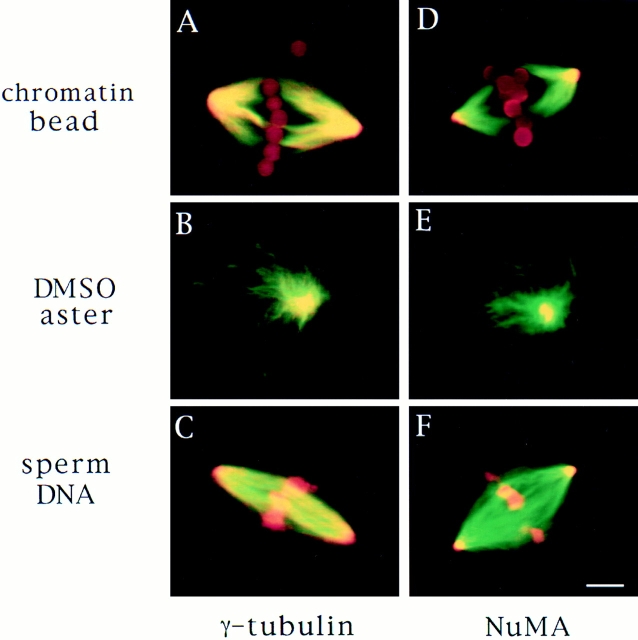
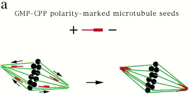
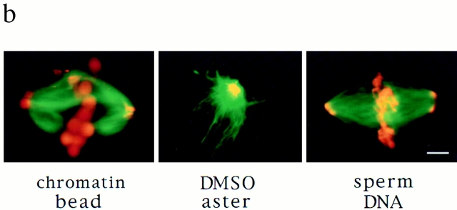
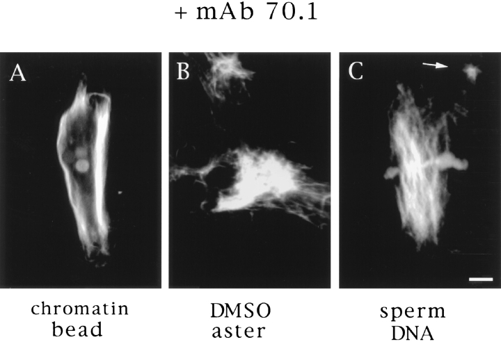
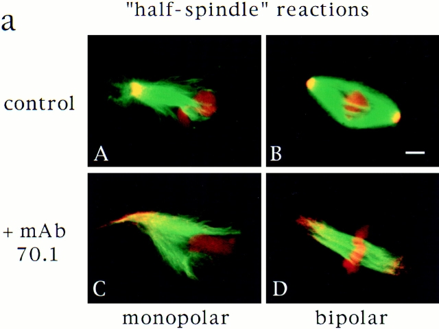
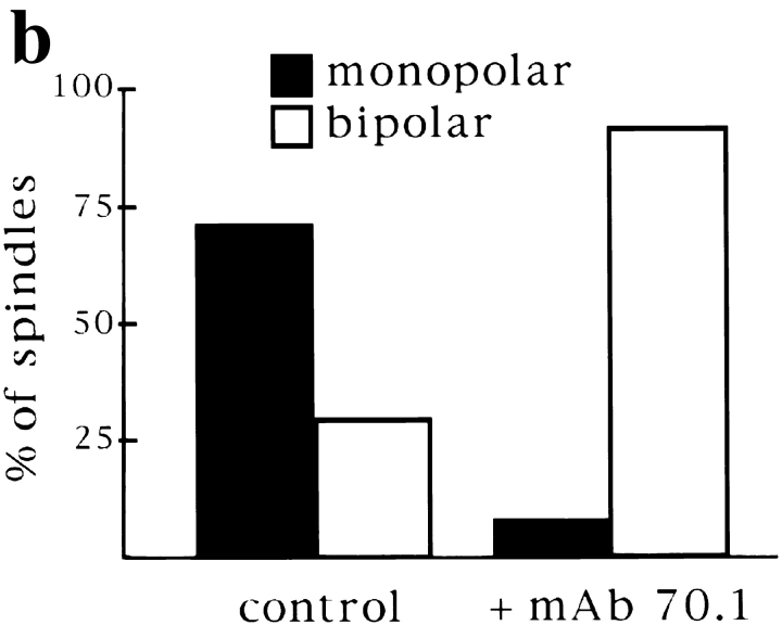
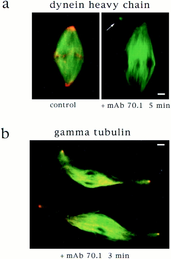
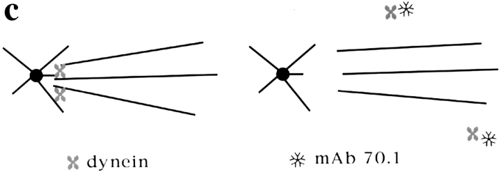
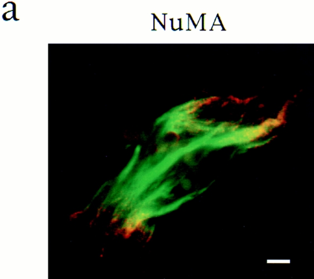
In Xenopus egg extracts, spindles assembled around sperm nuclei contain a centrosome at each pole, while those assembled around chromatin beads do not. Poles can also form in the absence of chromatin, after addition of a microtubule stabilizing agent to extracts. Using this system, we have asked (a) how are spindle poles formed, and (b) how does the nucleation and organization of microtubules by centrosomes influence spindle assembly? We have found that poles are morphologically similar regardless of their origin. In all cases, microtubule organization into poles requires minus end-directed translocation of microtubules by cytoplasmic dynein, which tethers centrosomes to spindle poles. However, in the absence of pole formation, microtubules are still sorted into an antiparallel array around mitotic chromatin. Therefore, other activities in addition to dynein must contribute to the polarized orientation of microtubules in spindles. When centrosomes are present, they provide dominant sites for pole formation. Thus, in Xenopus egg extracts, centrosomes are not necessarily required for spindle assembly but can regulate the organization of microtubules into a bipolar array.


Figures

Figure 1
Figure 1
Spindle pole antigens are similarly localized on poles whether or not centrosomes are present. Microtubules are green, antigens and DNA are red, and overlap is yellow. γ Tubulin (A–C) and NuMA (D–F) immunofluorescence of chromatin bead spindles (A and D) and DMSO asters (B and E) with self-assembled poles and sperm DNA spindles (C and F) with a centrosome at each pole. Bar, 5 μm.
Figure 2
Figure 2
Stable microtubule seeds translocate to and accumulate at mitotic poles in the presence and absence of centrosomes. Seeds are red, microtubules are green, and overlap is yellow. (a) Diagram of seed experiment: polarity-marked microtubules polymerized from pure tubulin with a nonhydrolyzable GTP analogue, GMP-CPP, were added to extracts containing spindles. Seeds bound to spindle microtubules and moved poleward, where they accumulated. (b) Seeds accumulate at poles of chromatin bead spindles, DMSO asters, and sperm DNA spindles that contain centrosomes. Bar, 5 μm.
Figure 2
Figure 2
Stable microtubule seeds translocate to and accumulate at mitotic poles in the presence and absence of centrosomes. Seeds are red, microtubules are green, and overlap is yellow. (a) Diagram of seed experiment: polarity-marked microtubules polymerized from pure tubulin with a nonhydrolyzable GTP analogue, GMP-CPP, were added to extracts containing spindles. Seeds bound to spindle microtubules and moved poleward, where they accumulated. (b) Seeds accumulate at poles of chromatin bead spindles, DMSO asters, and sperm DNA spindles that contain centrosomes. Bar, 5 μm.
Figure 3
Figure 3
Video analysis of seed movement on DMSO asters. (a) Successive video frames at 20-s intervals showing that individual seeds move poleward. (b) Rates of seed movement over 5-s intervals and distances of individual seeds from the pole over time. (c) Polarity-marked microtubule seeds are oriented with their minus ends directed toward the center of the aster and its focus of microtubule minus ends. Bars: (a) 5 μm; (c) 8 μm.
Figure 3
Figure 3
Video analysis of seed movement on DMSO asters. (a) Successive video frames at 20-s intervals showing that individual seeds move poleward. (b) Rates of seed movement over 5-s intervals and distances of individual seeds from the pole over time. (c) Polarity-marked microtubule seeds are oriented with their minus ends directed toward the center of the aster and its focus of microtubule minus ends. Bars: (a) 5 μm; (c) 8 μm.
Figure 4
Figure 4
Disruption of poles by antidynein antibodies. The monoclonal antibody (mAb 70.1) that recognizes the intermediate chain of cytoplasmic dynein was added to extracts containing chromatin bead spindles (A), DMSO asters (B), or sperm DNA spindles (C) with centrosomes. Pole structures were disrupted within 10 min. Bar, 5 μm.
Figure 8
Figure 8
Centrosomes are dominant sites of pole assembly. (a) Sperm half spindle reactions in the presence of control antibodies (A and B) or mAb 70.1 (C and D) were evaluated for microtubule polarity by NuMA immunofluorescence. (b) Quantification of monopolar and bipolar structures under each condition. At least 300 spindles were counted for each condition in two separate experiments. Bar, 5 μm.
Figure 8
Figure 8
Centrosomes are dominant sites of pole assembly. (a) Sperm half spindle reactions in the presence of control antibodies (A and B) or mAb 70.1 (C and D) were evaluated for microtubule polarity by NuMA immunofluorescence. (b) Quantification of monopolar and bipolar structures under each condition. At least 300 spindles were counted for each condition in two separate experiments. Bar, 5 μm.
Figure 5
Figure 5
Dynein tethers centrosomes to spindle poles. (a) Cytoplasmic dynein is eluted from spindles by addition of mAb 70.1. Immunofluorescent localization of dynein heavy chain to spindle poles disappears within 5 min after mAb 70.1 addition. Microtubules are green, dynein heavy chain is red, and overlap is yellow. (b) Centrosomes are released from sperm DNA spindle poles 3 min after addition of mAb 70.1. Immunofluorescent localization of γ tubulin on sperm centriolar structures that are dissociating from spindle poles. (c) Model of how dynein tethers spindle microtubules to centrosomal microtubules and how this is disrupted by mAb 70.1. Bars, 5 μm.
Figure 5
Figure 5
Dynein tethers centrosomes to spindle poles. (a) Cytoplasmic dynein is eluted from spindles by addition of mAb 70.1. Immunofluorescent localization of dynein heavy chain to spindle poles disappears within 5 min after mAb 70.1 addition. Microtubules are green, dynein heavy chain is red, and overlap is yellow. (b) Centrosomes are released from sperm DNA spindle poles 3 min after addition of mAb 70.1. Immunofluorescent localization of γ tubulin on sperm centriolar structures that are dissociating from spindle poles. (c) Model of how dynein tethers spindle microtubules to centrosomal microtubules and how this is disrupted by mAb 70.1. Bars, 5 μm.
Figure 6
Figure 6
Microtubules are sorted into antiparallel arrays around chromatin in the absence of pole formation. (a) Immunofluorescent localization of NuMA to the frayed ends of chromatin bead spindles that have formed in the absence of dynein activity. (b and c) Hooking analysis. (b) Quantification of hook handedness in control and poleless spindles. Percentage of right- and left-handed hooks in sections through spindle centers containing chromatin beads, and spindle ends containing microtubule poles (control), or bundles (+ mAb 70.1). (c) Low magnification micrographs (5-μm width) are shown to give an overall impression of microtubule organization seen in sections through spindle ends containing microtubule poles or bundles and through spindle centers containing beads. (d) Higher magnification micrographs (2–2.5-μm width) show hooks on cross sectioned individual microtubules. In the presence of control antibodies, a section through a pole contains right-handed (clockwise) hooks, while a section containing beads contains both right- and left-handed hooks. In the presence of mAb 70.1, a section through a microtubule bundle likely to be at the spindle end is shown that contains almost exclusively left-handed hooks. Note: Hook handedness does not give any information about the polarity of the microtubules in these sections but indicates the degree to which microtubule polarity is uniform. 50–100 microtubules were evaluated in each section, and three sections were evaluated for each condition. Bar, 5 μm.
Figure 6
Figure 6
Microtubules are sorted into antiparallel arrays around chromatin in the absence of pole formation. (a) Immunofluorescent localization of NuMA to the frayed ends of chromatin bead spindles that have formed in the absence of dynein activity. (b and c) Hooking analysis. (b) Quantification of hook handedness in control and poleless spindles. Percentage of right- and left-handed hooks in sections through spindle centers containing chromatin beads, and spindle ends containing microtubule poles (control), or bundles (+ mAb 70.1). (c) Low magnification micrographs (5-μm width) are shown to give an overall impression of microtubule organization seen in sections through spindle ends containing microtubule poles or bundles and through spindle centers containing beads. (d) Higher magnification micrographs (2–2.5-μm width) show hooks on cross sectioned individual microtubules. In the presence of control antibodies, a section through a pole contains right-handed (clockwise) hooks, while a section containing beads contains both right- and left-handed hooks. In the presence of mAb 70.1, a section through a microtubule bundle likely to be at the spindle end is shown that contains almost exclusively left-handed hooks. Note: Hook handedness does not give any information about the polarity of the microtubules in these sections but indicates the degree to which microtubule polarity is uniform. 50–100 microtubules were evaluated in each section, and three sections were evaluated for each condition. Bar, 5 μm.
Figure 7
Figure 7
Centrosomes provide permanent microtubule organizing sites. Sperm DNA and chromatin bead spindles before and 5 or 20 min after incubation on ice to depolymerize microtubules. No microtubules were visible on chromatin beads 5 min after cooling. Bar, 5 μm.

References

    1. Albertson DG, Thomson JN. Segregation of holocentric chromosomes at meiosis in the nematode, Caenorhabditis elegans. . Chromosome Res. 1993;1:15–26. - PubMed
    1. Bajer AS. Functional autonomy of monopolar spindle and evidence for oscillatory movement in mitosis. J Cell Biol. 1982;93:33–48. - PMC - PubMed
    1. Bajer AS, Mole BJ. Asters, poles, and transport properties within spindlelike microtubule arrays. Cold Spring Harbor Symp Quant Biol. 1982;1:263–283. - PubMed
    1. Bastmeyer M, Steffen W, Fuge H. Immunostaining of spindle components in tipulid spermatocytes using a serum against pericentriolar material. Eur J Cell Biol. 1986;42:305–310. - PubMed
    1. Belmont LD, Hyman AA, Sawin KE, Mitchison TJ. Real-time visualization of cell cycle-dependent changes in microtubule dynamics in cytoplasmic extracts. Cell. 1990;62:579–589. - PubMed