. 1997 Aug 11;138(3):707-17.
doi: 10.1083/jcb.138.3.707.

CD36 mediates the In vitro inhibitory effects of thrombospondin-1 on endothelial cells

Affiliations

CD36 mediates the In vitro inhibitory effects of thrombospondin-1 on endothelial cells

D W Dawson et al. J Cell Biol. .

Abstract

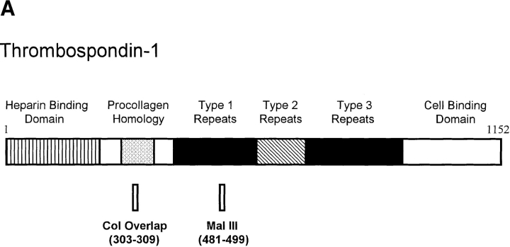
Thrombospondin-1 (TSP-1) is a naturally occurring inhibitor of angiogenesis that is able to make normal endothelial cells unresponsive to a wide variety of inducers. Here we use both native TSP-1 and small antiangiogenic peptides derived from it to show that this inhibition is mediated by CD36, a transmembrane glycoprotein found on microvascular endothelial cells. Both IgG antibodies against CD36 and glutathione-S-transferase-CD36 fusion proteins that contain the TSP-1 binding site blocked the ability of intact TSP-1 and its active peptides to inhibit the migration of cultured microvascular endothelial cells. In addition, antiangiogenic TSP-1 peptides inhibited the binding of native TSP-1 to solid phase CD36 and its fusion proteins, as well as to CD36-expressing cells. Additional molecules known to bind CD36, including the IgM anti-CD36 antibody SM, oxidized (but not unoxidized) low density lipoprotein, and human collagen 1, mimicked TSP-1 by inhibiting the migration of human microvascular endothelial cells. Transfection of CD36-deficient human umbilical vein endothelial cells with a CD36 expression plasmid caused them to become sensitive to TSP-1 inhibition of their migration and tube formation. This work demonstrates that endothelial CD36, previously thought to be involved only in adhesion and scavenging activities, may be essential for the inhibition of angiogenesis by thrombospondin-1.


Figures

Figure 1
Figure 1
The relationship of small TSP-1 peptides to the 180-kD monomer of TSP-1 and of GST–CD36 fusion proteins to the whole CD36 receptor protein. Numbers indicate amino acid residues present in the peptide or fusion protein. Shading on the whole CD36 molecule defines the minimal region of CD36 required for binding to TSP-1 (Pearce et al., 1995). Actual peptide sequences and detailed description of the fusion proteins are included in Materials and Methods.
Figure 1
Figure 1
The relationship of small TSP-1 peptides to the 180-kD monomer of TSP-1 and of GST–CD36 fusion proteins to the whole CD36 receptor protein. Numbers indicate amino acid residues present in the peptide or fusion protein. Shading on the whole CD36 molecule defines the minimal region of CD36 required for binding to TSP-1 (Pearce et al., 1995). Actual peptide sequences and detailed description of the fusion proteins are included in Materials and Methods.
Figure 2
Figure 2
Interference with the activity of TSP-1 and its antiangiogenic peptides by soluble GST–CD36 fusion proteins. Increasing concentrations of CD36 fusion proteins that contain a TSP-1 binding site, FP93-120 (circles) or FP93-298 (triangles), or a CD36 fusion protein that lacks a TSP-1 binding site, FP298-439 (squares), were preincubated for 2 h at 4°C with (A) 2 nM TSP-1, (B) 10 μM Mal III peptide, (C) 30 μM Col overlap peptide, or (D) control media. Each mixture was then tested for the ability to block bovine capillary endothelial cell migration towards bFGF (solid symbols) or influence background migration in the absence of bFGF (D, open symbols). Data, accumulated from nine experiments, are reported as a percentage of maximum migration, where 100% represents the number of cells migrating towards the inducer bFGF alone, and 0% corresponds to the number of cells migrating randomly in the absence of inducer (Bkgd). 100% varied between experiments from 32 to 81 cells migrated/10 high-power fields. Bars indicate standard errors.
Figure 2
Figure 2
Interference with the activity of TSP-1 and its antiangiogenic peptides by soluble GST–CD36 fusion proteins. Increasing concentrations of CD36 fusion proteins that contain a TSP-1 binding site, FP93-120 (circles) or FP93-298 (triangles), or a CD36 fusion protein that lacks a TSP-1 binding site, FP298-439 (squares), were preincubated for 2 h at 4°C with (A) 2 nM TSP-1, (B) 10 μM Mal III peptide, (C) 30 μM Col overlap peptide, or (D) control media. Each mixture was then tested for the ability to block bovine capillary endothelial cell migration towards bFGF (solid symbols) or influence background migration in the absence of bFGF (D, open symbols). Data, accumulated from nine experiments, are reported as a percentage of maximum migration, where 100% represents the number of cells migrating towards the inducer bFGF alone, and 0% corresponds to the number of cells migrating randomly in the absence of inducer (Bkgd). 100% varied between experiments from 32 to 81 cells migrated/10 high-power fields. Bars indicate standard errors.
Figure 3
Figure 3
Inhibition of TSP-1 binding to CD36 and its fusion proteins by antiangiogenic TSP-1 peptides. (A) Binding of 125I–TSP-1 (20 μg/ ml) to a confluent monolayer of Bowes melanoma cells expressing CD36 was determined in the presence of increasing concentrations of antiangiogenic TSP-1 peptides Mal III (circles, lowest curve), Mal III variant (triangles, second lowest curve), and Col overlap (squares, third lowest curve) or of control peptide LYPQHKT (diamonds, top curve). Data were normalized as a percentage of TSP-1 bound under control conditions. (B) The effects of TSP-1 peptides Mal III and Col overlap on the binding of 125I–TSP-1 (20 μg/ml) to solid phase CD36 and CD36 fusion proteins FP67-157 and FP93-120, which contain a TSP-1 binding site, and to FP298-439, which does not, are shown. Nonspecific binding was determined in the presence of 5 mM EDTA. Bars indicate standard error (n = 3 for A and n = 5 for B).
Figure 3
Figure 3
Inhibition of TSP-1 binding to CD36 and its fusion proteins by antiangiogenic TSP-1 peptides. (A) Binding of 125I–TSP-1 (20 μg/ ml) to a confluent monolayer of Bowes melanoma cells expressing CD36 was determined in the presence of increasing concentrations of antiangiogenic TSP-1 peptides Mal III (circles, lowest curve), Mal III variant (triangles, second lowest curve), and Col overlap (squares, third lowest curve) or of control peptide LYPQHKT (diamonds, top curve). Data were normalized as a percentage of TSP-1 bound under control conditions. (B) The effects of TSP-1 peptides Mal III and Col overlap on the binding of 125I–TSP-1 (20 μg/ml) to solid phase CD36 and CD36 fusion proteins FP67-157 and FP93-120, which contain a TSP-1 binding site, and to FP298-439, which does not, are shown. Nonspecific binding was determined in the presence of 5 mM EDTA. Bars indicate standard error (n = 3 for A and n = 5 for B).
Figure 4
Figure 4
Interference with the activity of TSP-1 and its antiangiogenic peptides by monoclonal IgG antibodies against CD36. Monoclonal IgG antibodies against CD36, FA6-152, and OKM-5 or an isotype-matched control were tested at 10 μg/ml for ability to block the inhibition of human microvascular endothelial cell migration towards bFGF by (A) TSP-1 (2 nM) or (B) TSP-1 peptides Mal III (30 μM) or Col overlap (50 μM). Angiostatin (2 μg/ml) served as a control inhibitor. Data from three separate experiments were normalized and reported as in Fig. 1. 100% varied between experiments from 32 to 53 cells migrated/10 high-power fields. *Samples significantly different from parallel condition using control media, P < 0.02.
Figure 4
Figure 4
Interference with the activity of TSP-1 and its antiangiogenic peptides by monoclonal IgG antibodies against CD36. Monoclonal IgG antibodies against CD36, FA6-152, and OKM-5 or an isotype-matched control were tested at 10 μg/ml for ability to block the inhibition of human microvascular endothelial cell migration towards bFGF by (A) TSP-1 (2 nM) or (B) TSP-1 peptides Mal III (30 μM) or Col overlap (50 μM). Angiostatin (2 μg/ml) served as a control inhibitor. Data from three separate experiments were normalized and reported as in Fig. 1. 100% varied between experiments from 32 to 53 cells migrated/10 high-power fields. *Samples significantly different from parallel condition using control media, P < 0.02.
Figure 5
Figure 5
Inhibition of endothelial cell migration by additional CD36 ligands. (A) A murine anti-CD36 IgM monoclonal antibody SM∅ and an isotype-matched control monoclonal antibody were tested at 1 μg/ml for ability to inhibit human microvascular endothelial cell migration towards bFGF. Data were normalized and reported as in Fig. 1. *Significant inhibition compared with bFGF tested alone, P < 0.001. (B) Human collagen I, LDL, and OxLDL were tested at 2 μg/ml in the presence or absence of 10 μg/ml of monoclonal antibodies against CD36 for ability to inhibit migration of human microvascular cells. Data from two experiments were normalized and reported as in Fig 1. When tested alone, collagen I, OxLDL, and LDL had no significant effect on migration. 100% varied between experiments from 29 to 42 cells migrated/10 high-power fields. *Samples significantly different from bFGF tested with control media, P < 0.001.
Figure 5
Figure 5
Inhibition of endothelial cell migration by additional CD36 ligands. (A) A murine anti-CD36 IgM monoclonal antibody SM∅ and an isotype-matched control monoclonal antibody were tested at 1 μg/ml for ability to inhibit human microvascular endothelial cell migration towards bFGF. Data were normalized and reported as in Fig. 1. *Significant inhibition compared with bFGF tested alone, P < 0.001. (B) Human collagen I, LDL, and OxLDL were tested at 2 μg/ml in the presence or absence of 10 μg/ml of monoclonal antibodies against CD36 for ability to inhibit migration of human microvascular cells. Data from two experiments were normalized and reported as in Fig 1. When tested alone, collagen I, OxLDL, and LDL had no significant effect on migration. 100% varied between experiments from 29 to 42 cells migrated/10 high-power fields. *Samples significantly different from bFGF tested with control media, P < 0.001.
Figure 6
Figure 6
Sensitivity to TSP-1 inhibition of tube formation induced by CD36 expression in CD36-deficient HUVECs. The first column shows FACS® analysis for CD36 expression performed on untransfected HUVECs, a transfectant expressing very low levels of CD36 (clone 35), and two transfectants expressing higher levels of CD36 (clones 31 and 36). Fluorescence is plotted vs. cell number. The dotted line in the clones reproduces the HUVEC CD36 null pattern. The morphology of each of the transfectants grown in two-dimensional cell culture is shown in the second column. In the third column, tube formation in a three-dimensional culture of Matrigel is pictured for each line.
Figure 7
Figure 7
Sensitivity to TSP-1 inhibition of migration after transfection of HUVECs with CD36. Clones described in Fig. 5 were tested for sensitivity to inhibition of migration by 2 nM TSP-1 in the presence and absence of 10 μg/ml anti-CD36 monoclonal antibody OKM-5. Data from four separate experiments were normalized and reported as in Fig 1. 100% varied between experiments from 37 to 82 cells migrated/10 high-power fields. *Samples that differ significantly from bFGF tested alone, P < 0.02.

References

    1. Asch AS, Barnwell J, Silverstein RL, Nachman RL. Isolation of the thrombospondin membrane receptor. J Clin Invest. 1987;79:1054–1061. - PMC - PubMed
    1. Asch AS, Silbiger S, Heimer E, Nachman RL. Thrombospondin sequence motif (CSVTCG) is responsible for CD36 binding. Biochem Biophys Res Commun. 1992;182:1208–1217. - PubMed
    1. Asch AS, Liu I, Briccetti FM, Barnwell JW, Kwakye-Berko F, Dokun A, Goldberger J, Pernambuco M. Analysis of CD36 binding domains: ligand specificity controlled by dephosphorylation of an ectodomain. Science (Wash DC) 1993;262:1436–1440. - PubMed
    1. Bagavandoss P, Wilkes JW. Specific inhibition of endothelial cell proliferation by thrombospondin. Biochem Biophys Res Commun. 1990;170:867–872. - PubMed
    1. Baruch DI, Gormely JA, Ma C, Howard RJ, Pasloske BL. Plasmodium falciparum erythrocyte membrane protein 1 is a parasitized erythrocyte receptor for adherence to CD36, thrombospondin, and intracellular adhesion molecule 1. Proc Natl Acad Sci USA. 1996;93:3497–3502. - PMC - PubMed

Publication types

MeSH terms