. 1997 Aug 11;138(3):719-28.
doi: 10.1083/jcb.138.3.719.

Laminin 5 binds the NC-1 domain of type VII collagen

Affiliations

Laminin 5 binds the NC-1 domain of type VII collagen

P Rousselle et al. J Cell Biol. .

Abstract

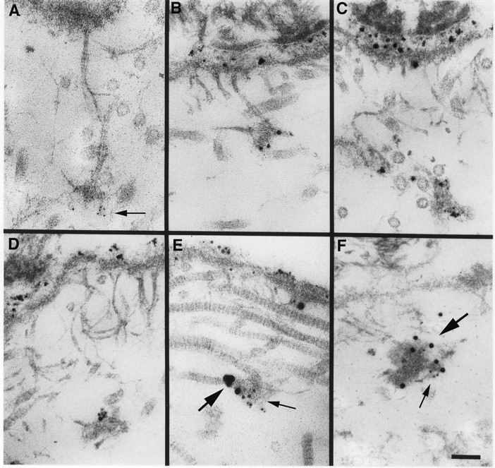
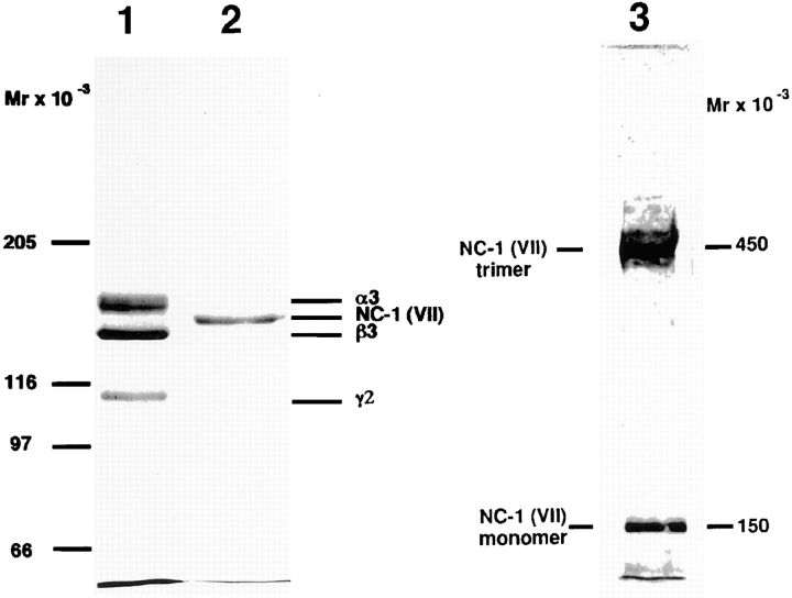
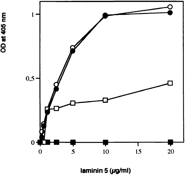
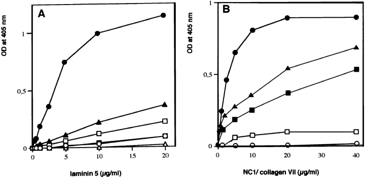
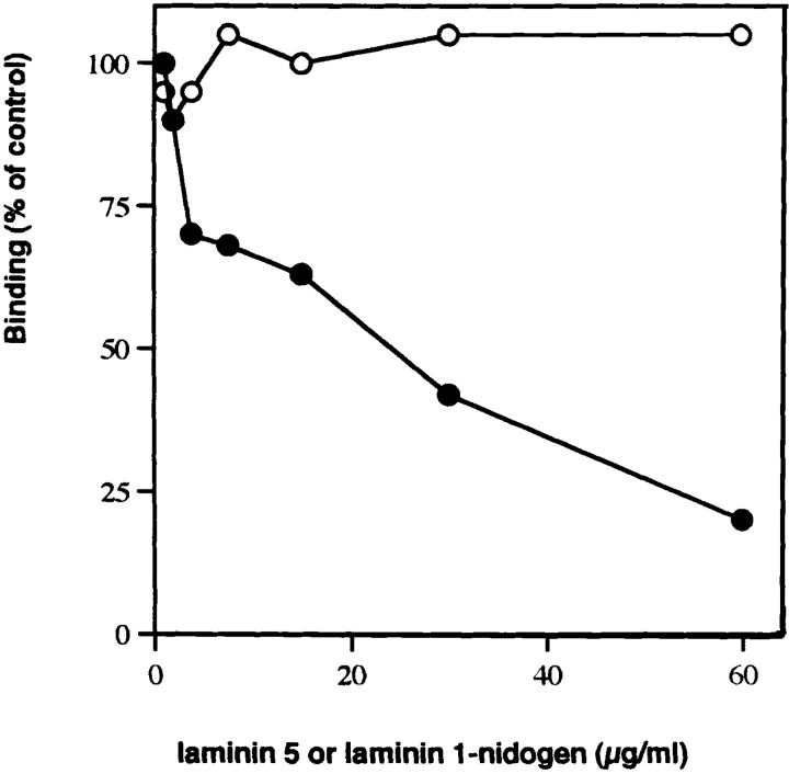
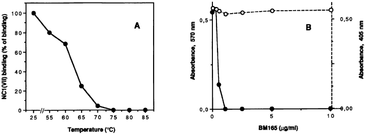
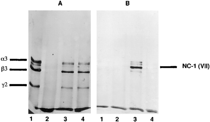
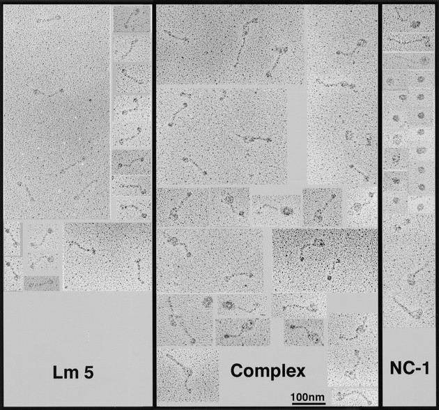
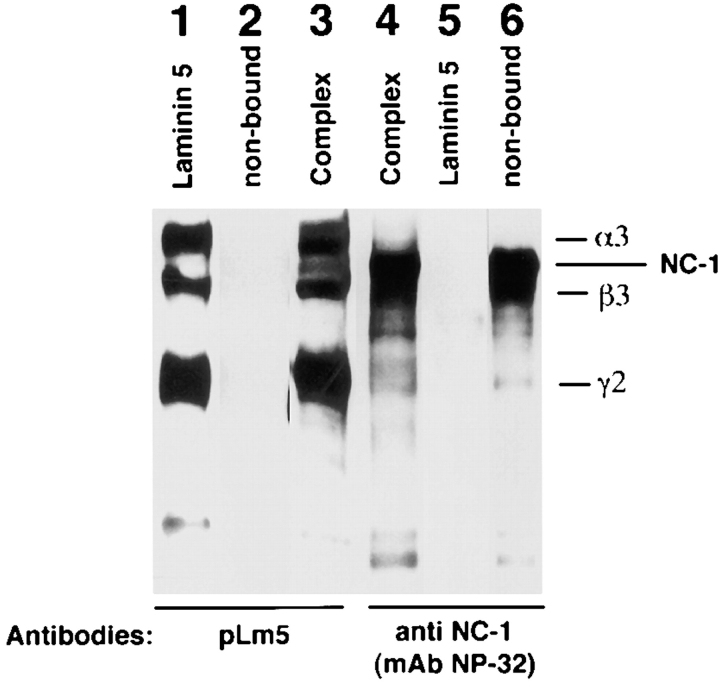
Mutational analyses of genes that encode components of the anchoring complex underlying the basolateral surface of external epithelia indicate that this structure is the major element providing for resistance to external friction. Ultrastructurally, laminin 5 (alpha3beta3gamma2; a component of the anchoring filament) appears as a thin filament bridging the hemidesmosome with the anchoring fibrils. Laminin 5 binds the cell surface through hemidesmosomal integrin alpha6beta4. However, the interaction of laminin 5 with the anchoring fibril (type VII collagen) has not been elucidated. In this study we demonstrate that monomeric laminin 5 binds the NH2-terminal NC-1 domain of type VII collagen. The binding is dependent upon the native conformation of both laminin 5 and type VII collagen NC-1. Laminin 6 (alpha3beta1gamma1) has no detectable affinity for type VII collagen NC-1, indicating that the binding is mediated by the beta3 and/or gamma2 chains of laminin 5. Approximately half of the laminin 5 solubilized from human amnion or skin is covalently complexed with laminins 6 or 7 (alpha3beta2gamma1). The adduction occurs between the NH2 terminus of laminin 5 and the branch point of the short arms of laminins 6 or 7. The results are consistent with the presumed orientation of laminin 5, having the COOH-terminal G domain apposed to the hemidesmosomal integrin, and the NH2-terminal domains within the lamina densa. The results also support a model predicting that monomeric laminin 5 constitutes the anchoring filaments and bridges integrin alpha6beta4 with type VII collagen, and the laminin 5-6/7 complexes are present within the interhemidesmosomal spaces bound at least by integrin alpha3beta1 where they may mediate basement membrane assembly or stability, but contribute less significantly to epithelial friction resistance.


Figures

Figure 9
Figure 9
Localization of laminin 5 to anchoring plaques. Laminin 5 is localized to anchoring plaques in human skin using mAb BM165 and 5-nm (A, arrow) or 1 nm silver-enhanced colloidal gold (B–D). Labeling of the lamina lucida and anchoring plaques is intense using silver-enhanced 1 nm colloidal gold in comparison to 5 nm colloidal gold. Using a mixture of mAb BM165 (anti-laminin α3) and pAb 3959 (anti-type VII collagen NC-1), laminin 5 is seen to colocalize with type VII collagen in anchoring plaques (E, 1-nm silver-enhanced particles specific for mAb BM165 [small arrow] and 10-nm silver-enhanced particles are specific for pAb 3959 [large arrow]; F, 5-nm particles specific for mAb BM165 (small arrow) and 15-nm particles specific for pAb 3959 [large arrow]. Bar, 100 nm.
Figure 1
Figure 1
Electrophoretic analysis of immunoaffinity-purified laminin 5 from cell culture media and type VII collagen–NC-1 from collagenase extracts of amniotic membranes. 2 μg of materials affinity purified on mAb 6/F12 from SCC25 cell culture medium (lane 1), and 2 μg of materials affinity purified on mAb NP-32 from collagenase extracts of amniotic membranes (lane 2) were separated by SDS-PAGE after reduction on a 3–7.5% gradient polyacrylamide gel and visualized by Coomassie blue staining. Consistent with previous results, the reduced bands representing subunits of laminin 5 (α3, 165 kD; β3, 140 kD; γ2, 155 and 105 kD) and type VII collagen–NC-1 (150 kD). The electrophoretic migration position of purified NC-1 before disulfide-bond reduction is shown in lane 3. Migration positions of the molecular weight markers are indicated to the left of each gel.
Figure 2
Figure 2
Binding of soluble laminin 5 to immobilized type VII procollagen. Microtiter wells were coated with 1 μg of type VII–NC-1 purified from amnion (•) or intact type VII procollagen immunopurified from WISH cell culture medium (□), or type VII procollagen digested with either bacterial collagenase (○) or pepsin (▪). pAb to laminin 5 were used for detecting the amount of bound substrate.
Figure 3
Figure 3
(A) Interaction of laminin 5 with various immobilized extracellular matrix components Binding of soluble laminin 5 to immobilized collagen I (□), collagen IV (▪), collagen XII (▴), collagen XIV (▵), NC-1-VII (•), and laminin 1–nidogen complex (○) at a concentration of 1 μg per well was determined by a pAb in an enzyme-coupled second antibody reaction as described in Materials and Methods. (B) Interaction of type VII–NC-1 with type IV collagen and with various laminin isoforms. Binding of soluble type VII–NC-1 to immobilized collagen IV (▪), laminin 1–nidogen complex (○), laminin 5 (•), laminin 6 (□), and laminin 5–laminin 6 complex (▴) at the concentration of 1 μg per well was determined by a pAb in an enzyme-coupled second antibody reaction as described in Materials and Methods.
Figure 4
Figure 4
Inhibition of type VII–NC-1 binding to immobilized laminin 5 by preincubation with various competitors. The test system consisted of laminin 5 as the immobilized phase at the concentration of 1 μg per well, and a fixed amount of soluble type VII collagen– NC-1 (5 μg/ml). Type VII– NC1 was incubated at 4°C for 8 h with varying concentration of laminin 5 (•) or laminin 1–nidogen complex (○) before testing on the insoluble ligand.
Figure 5
Figure 5
(A) Effect of heat denaturation on laminin 5 interaction with type VII–NC-1. Multiwell plates were coated with laminin 5 (•) at concentration of 1 μg before or after heating at the indicated temperatures. Soluble type VII–NC-1 was then added at concentration of 10 μg/ml and bound material was determined by a pAb in an enzyme-coupled second antibody reaction as described in Materials and Methods. (B) Effects of mAb BM165 on adhesion of normal human keratinocytes and on binding of type VII–NC-1 to laminin 5. Multiwell plates were coated with laminin 5 at 10 μg/ml. After saturation, the wells were incubated with indicated concentrations of mAb BM165 for 60 min before the cell adhesion experiment (•), or together with soluble type VII–NC-1 (10 μg/ml) for the binding assay (○). Extent of cell adhesion was measured with a colorimetric reaction at 570 nm. Each point represents the average of triplicate wells. In the ELISA assay, bound type VII–NC-1 was determined by a pAb in an enzyme-coupled second antibody reaction as described in Materials and Methods. Because the OD values in both systems were in the same range, the data were presented on a single graph.
Figure 6
Figure 6
Western blot analysis of the complex of laminin 5 and NC-1 produced in solution. Laminin 5 alone (lanes A1 and B1), NC-1 alone (lanes A2 and B2), a mixture of laminin 5 and NC-1 (lanes A3 and B3), or a mixture of laminin 5 and heat denatured NC-1 (lanes A4 and B4) were immunoprecipitated with anti-α3 mAb (BM-165). The contents of the precipitates were separated by SDS-PAGE after disulfide bond reduction and visualized by Western analysis using (A) polyclonal anti-laminin 5 or (B) monoclonal anti-NC-1 (mAb NP-32).
Figure 7
Figure 7
Visualization of the laminin 5–NC-1 complex formed in solution by transmission electron microscopy after rotary shadowing. Collagen VII–NC-1, laminin 5, and a mixture of both molecules were dialyzed against 0.2 M ammonium bicarbonate, diluted 1:1 with glycerol, and rotary shadowed. (Lm5) The images visualized are characteristic of laminin 5 molecules. The 107-nm molecules appear as extended rods with globular knobs at each terminus. (NC1) Under the conditions used to maintain the stability of the complex, NC-1 appears as an asymmetrical spheroid, ∼41 nm in narrowest diameter. Due to incomplete digestion, helical domains of type VII collagen can be seen on some NC-1 molecules. (Complex) Images of the laminin 5–type VII collagen–NC-1 complex preparation contains uncomplexed laminin 5 and NC-1, and images that appear as a complex of one laminin 5 molecule with one NC-1 molecule, in which the long arm may correspond to laminin 5 and the big globular domain to NC-1.
Figure 8
Figure 8
Western blot analysis of the laminin 5–type VII collagen–NC-1 complex immunoaffinity purified from collagenase extracts of amniotic membranes. Type VII collagen–NC-1 was first affinity purified on mAb NP-32 from collagenase extracts of amniotic membranes. Eluted fractions of NC-1 were pooled, dialyzed against PBS, and passed over a mAb 6F12 affinity chromatography column. Bound materials were immunoblotted with polyclonal anti-laminin 5 (pLm5; lane 3), and anti-NC-1 (mAb NP-32, lane 4). The bound material shows the pattern obtained for purified laminin 5 (lane 1), which does not contain NC-1 (lane 2) and for purified NC-1 (not shown, but identical to lane 6). The nonbound fraction contains NC-1 only (lane 6) but no laminin 5 (lane 2).
Figure 10
Figure 10
Model for the presence of monomeric laminin 5 and complexed laminin 5 within an epithelial–stromal junction. The cartoon depicts monomeric laminin 5 as the bridge between hemidesmosomal integrin α6β4 and the type VII collagen NC-1 domain. The tight binding of laminin 5 to α6β4 and to type VII collagen provides the primary resistance to frictional forces. The laminin 5–6/7 complex is shown within the basement membrane between hemidesmosomes, bound by integrin α3β1 where it potentially functions to maintain basement membrane stability and contact with the epithelial basolateral surface.

References

    1. Aubert-Foucher E, Font B, Eichenberger D, Goldschmidt D, Lethias C, van der Rest M. Purification and partial characterization of native type XIV collagen. J Biol Chem. 1992;267:15759–15764. - PubMed
    1. Aumailley M, Wiedemann H, Mann K, Timpl R. Binding of nidogen and the laminin-nidogen complex to basement membrane collagen type IV. Eur J Biochem. 1989;184:241–248. - PubMed
    1. Bachinger HP, Morris NP, Lunstrum GP, Keene DR, Rosenbaum LM, Compton LA, Burgeson RE. The relationship of the biophysical and biochemical characteristics of type VII collagen to the function of anchoring fibrils. J Biol Chem. 1990;265:10095–10101. - PubMed
    1. Bentz H, Morris NP, Murray L, Sakai LY, Hollister DW, Burgeson RE. Isolation and partial characterization of a new human collagen with an extended triple-helical structural domain. Proc Natl Acad Sci USA. 1983;80:3168–3172. - PMC - PubMed
    1. Bruckner-Tuderman L. Collagen VII and bullous disorders of the skin. Dermatology. 1994;189:16–20. - PubMed

Publication types