

An approximately 1.2-Mb bacterial artificial chromosome contig refines the genetic and physical maps of the lurcher locus on mouse chromosome 6
- PMID: 9253602
- PMCID: PMC310680
- DOI: 10.1101/gr.7.7.736
An approximately 1.2-Mb bacterial artificial chromosome contig refines the genetic and physical maps of the lurcher locus on mouse chromosome 6
Abstract
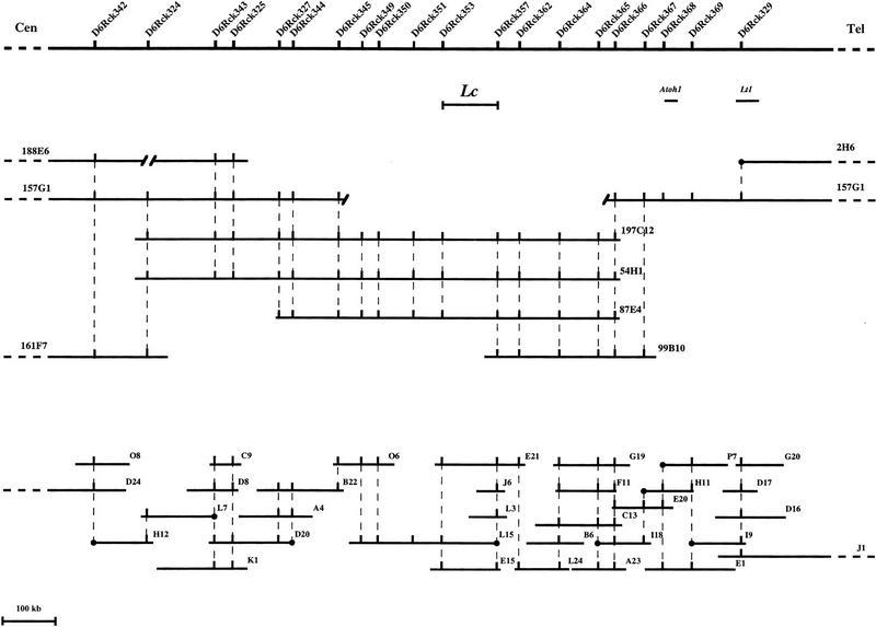
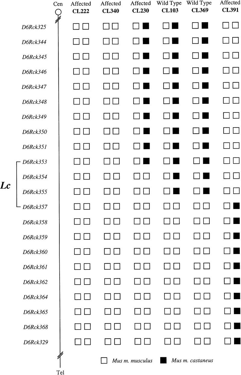
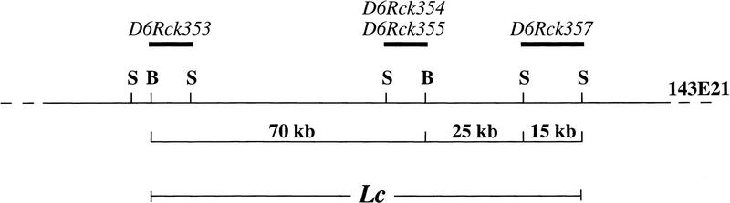
Lurcher (Lc) is a semidominant mouse mutant that displays a characteristic ataxia in the heterozygous state beginning in the third postnatal week. This symptom results from a neurodegenerative event in the cerebellum: There is a catastrophic loss of Purkinje cells in the heterozygote animal between postnatal days 10 and 15. In an effort to identify the genetic lesion borne by Lc mice, we initiated a cloning project based on the position of the Lc mutation on mouse chromosome 6. We have extended our previous analysis of the genomic segment containing the Lc locus by isolating a set of stable and manipulable genomic clones called bacterial artificial chromosomes (BACs) that cover this region of mouse chromosome 6. These clones provided a good substrate for the isolation of markers that were used to refine the physical map of the locus. Furthermore, 20 of these markers were mapped onto our (B6CBACa-AW-J/A-Lc x CAST/Ei)F1 x B6CBACa-AW-J/A backcross, refining the genetic map and identifying two nonrecombinant markers (D6Rck354 and D6Rck355). These two markers, in conjunction with the closest flanking markers, were used to identify a 110-kb genomic segment that contains all four markers and hence contains the Lc locus. This small genomic segment, covered by multiple BACs, sets the stage for the final effort of this project-the identification of transcripts and of the mutation within the Lc locus.
Figures
Similar articles
-
A new allele of the lurcher gene, lurcherJ.Mamm Genome. 1997 Sep;8(9):647-50. doi: 10.1007/s003359900530. Mamm Genome. 1997. PMID: 9271665
-
Generation of a high-resolution genetic map and a YAC contig of the Lurcher locus on mouse chromosome 6.Genome Res. 1995 Nov;5(4):381-92. doi: 10.1101/gr.5.4.381. Genome Res. 1995. PMID: 8750197
-
Genetic mapping of the lurcher locus on mouse chromosome 6 using an intersubspecific backcross.Genomics. 1991 Jan;9(1):147-53. doi: 10.1016/0888-7543(91)90232-4. Genomics. 1991. PMID: 1672287
-
Comparative mapping of distal murine chromosome 11 and human 17q21.3 in a region containing a modifying locus for murine plasma von Willebrand factor level.Genomics. 1998 Nov 15;54(1):19-30. doi: 10.1006/geno.1998.5553. Genomics. 1998. PMID: 9806826
-
Genetic and physical delineation of the region overlapping the progressive motor neuropathy (pmn) locus on mouse chromosome 13.Genomics. 2001 Jul;75(1-3):9-16. doi: 10.1006/geno.2001.6595. Genomics. 2001. PMID: 11472062
Cited by
-
The lurcher mutation and ionotropic glutamate receptors: contributions to programmed neuronal death in vivo.Brain Pathol. 1998 Oct;8(4):795-807. doi: 10.1111/j.1750-3639.1998.tb00201.x. Brain Pathol. 1998. PMID: 9804384 Free PMC article. Review.
-
A new allele of the lurcher gene, lurcherJ.Mamm Genome. 1997 Sep;8(9):647-50. doi: 10.1007/s003359900530. Mamm Genome. 1997. PMID: 9271665
References
-
- Akazawa C, Ishibashi M, Shimizu C, Nakanishi S, Kageyama R. A mammalian helix-loop-helix factor structurally related to the product of Drosophila proneural gene atonal is a positive transcriptional regulator expressed in the developing nervous system. J Biol Chem. 1995;270:8730–8738. - PubMed
-
- Bahary N, Pachter J E, Felman R, Leibel R L, Albright K, Cram S, Friedman J M. Molecular mapping of mouse Chromosomes 4 and 6: Use of a flow-sorted Robertsonian chromosome. Genomics. 1992;13:761–769. - PubMed
-
- Ben-Arie N, McCall AE, Berkman S, Eichele G, Bellen HJ, Zoghbi HY. Evolutionary conservation of sequence and expression of the bHLH protein Atonal suggests a conserved role in neurogenesis. Hum Mol Genet. 1996;5:1207–1216. - PubMed
-
- Caddy KW, Biscoe TJ. Structural and quantitative studies on the normal C3H and lurcher mutant mouse. [Review] Phil Trans Roy Soc Lond Ser B Biol Sci. 1979;287:167–201. - PubMed
Publication types
MeSH terms
Associated data
- Actions
- Actions
Grants and funding
LinkOut - more resources
Full Text Sources
Molecular Biology Databases
Miscellaneous