. 1997 Aug 18;186(4):479-87.
doi: 10.1084/jem.186.4.479.

Introduction of a glycosylation site into a secreted protein provides evidence for an alternative antigen processing pathway: transport of precursors of major histocompatibility complex class I-restricted peptides from the endoplasmic reticulum to the cytosol

Affiliations

Introduction of a glycosylation site into a secreted protein provides evidence for an alternative antigen processing pathway: transport of precursors of major histocompatibility complex class I-restricted peptides from the endoplasmic reticulum to the cytosol

I Bacik et al. J Exp Med. .

Abstract

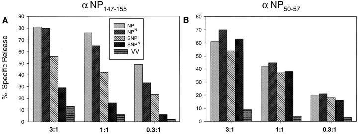
We found that the presentation of a H-2Kd-restricted determinant from influenza virus nucleoprotein (NP) to T cells is strictly dependent on expression of the transporter associated with antigen presentation (TAP), regardless of whether NP is expressed as a cytosolic or secreted NP (SNP). Introducing an N-linked glycosylation site into the determinant selectively reduced presentation of SNP. This indicates that glycosylation does not interfere with TAP-transported peptides, and therefore that cytosolic peptides derived from SNP must have been exposed to the glycosylation machinery of the endoplasmic reticulum (ER) before their existence in the cytosol. Based on these findings, we propose that TAP-dependent processing of at least some ER-targeted proteins entails the reimportation of protein from the secretory pathway to the cytosol, where the protein is processed via the classical pathway.


Figures

Figure 1
Figure 1
Peptide binding to Kd. RMA/S Kd cells incubated overnight at 26°C were incubated for an additional hour with the indicated concentration of peptide and BFA (5 μg/ml), and then at 37°C for 2 h (left). Alternatively, cells were incubated with 10 μM peptide and BFA, extensively washed, and then incubated at 37°C for the indicated time in the absence of peptide (right). The amount of cell-surface Kd was determined using the fluorescein-conjugated mAb SF1.1.1 (PharMingen, San Diego, CA). The concentration of peptide required for protection of one half of the Kd molecules was determined by linear extrapolation of the mean channel fluorescence values.
Figure 3
Figure 3
Antigenicity and immunogenicity of rVVs expressing various forms of NP. (A) L-Kd cells were infected for the indicated times with the rVVs indicated before BFA was added and then maintained continuously. After 51Cr labeling, cells were incubated for 6 h at an E/T of 5:1 with a TCD8+ line produced by PR8 in vitro secondary restimulation of splenocytes derived from mice inoculated with VV-NP. The recognition of synthetic peptides incubated with cells at the indicated concentration is shown in the inset. (B) Splenocytes from BALB/c mice immunized with the indicated rVV 3 wk previously were restimulated in vitro for 7 d with the homologous synthetic peptide (TYQRTRALV or TYNRTRALV), and tested for their ability to lyse P815 cells sensitized with the same peptide. Cultures were used at the same dilution. All values have been corrected for background lysis values obtained with cells in the absence of added synthetic peptides.
Figure 5
Figure 5
TAP dependence of antigen presentation. (A) T2 Kd cells coinfected with VV-TAP (1 and 2) and the rVVs indicated, or just with the rVV indicated (“No TAP”) were tested for lysis by a NP-specific TCD8+ line at the E/T ratios indicated. The recognition of synthetic peptides incubated with cells at the indicated concentration by the same TCD8+ line is shown in the inset. The amount of antigen expressed by the target cells 4 h after infection was determined by quantitating bands detected in SDS-PAGE of whole cell extracts as described in Fig. 2. The adjusted amounts of various forms of NP synthesized relative to NP (= 1.00) are: NPN, 1.49; SNP, 1.01; and SNPN, 1.01. (B) Effect of ICP47 on antigen presentation. T2 Kd cells infected with the rVVs indicated were tested for lysis by the TCD8+ line used in A at the indicated E/T ratio. (C) The target cells used in A were incubated in flat-bottom 96-well plates coated with Con A to adhere the cells. At the conclusion of the 51Cr release assay, cells were fixed by 2-min incubation with acetone/methanol (1:1) and indirectly immunoperoxidase stained using the H16-L10 mAb. Video images of the stained cells were printed using a printer (UP5500; Sony, San Jose, CA), digitized by a flat bed scanner, assembled using Adobe Photoshop software, and, printed with a digital printer (3000; Fuji Photo Film USA).
Figure 5
Figure 5
TAP dependence of antigen presentation. (A) T2 Kd cells coinfected with VV-TAP (1 and 2) and the rVVs indicated, or just with the rVV indicated (“No TAP”) were tested for lysis by a NP-specific TCD8+ line at the E/T ratios indicated. The recognition of synthetic peptides incubated with cells at the indicated concentration by the same TCD8+ line is shown in the inset. The amount of antigen expressed by the target cells 4 h after infection was determined by quantitating bands detected in SDS-PAGE of whole cell extracts as described in Fig. 2. The adjusted amounts of various forms of NP synthesized relative to NP (= 1.00) are: NPN, 1.49; SNP, 1.01; and SNPN, 1.01. (B) Effect of ICP47 on antigen presentation. T2 Kd cells infected with the rVVs indicated were tested for lysis by the TCD8+ line used in A at the indicated E/T ratio. (C) The target cells used in A were incubated in flat-bottom 96-well plates coated with Con A to adhere the cells. At the conclusion of the 51Cr release assay, cells were fixed by 2-min incubation with acetone/methanol (1:1) and indirectly immunoperoxidase stained using the H16-L10 mAb. Video images of the stained cells were printed using a printer (UP5500; Sony, San Jose, CA), digitized by a flat bed scanner, assembled using Adobe Photoshop software, and, printed with a digital printer (3000; Fuji Photo Film USA).
Figure 2
Figure 2
Characterization of SNP. (A) Aliquots of the target cells used in Fig. 3 A were removed 90 min after infection and radiolabeled with [35S]methionine. Total cell extracts were analyzed by SDS-PAGE. Radioactivity in the fixed and dried gels was located using a Phosphorimager that was used to quantitate the radioactivity in the NP and SNP band, which are clearly evident. The total amounts of counts per lane were normalized to allow for differences in sample preparation, and the corrected amount of radioactivity present in the same region of an extract prepared from cells infected with a control VV was subtracted from each value. The adjusted amounts of various forms of NP synthesized relative to NP (= 1.00) are: NPN, 1.65; NPNRA, 2.16; SNP, 1.24; SNPN, 1.72; SNPN high, 2.23; and SNPNRA, 1.82. (B) NP present in detergent lysates from cells infected with rVV expressing various forms of NP was collected using protein A agarose preloaded with the NP-specific mAb H16-L10, digested with endo H and analyzed by SDS-PAGE.
Figure 4
Figure 4
Comparison of presentation of NP147-155 to NP50-57. L-Kd cells infected with the rVV indicated were tested for lysis by TCD8+ lines specific for NP147-155 or NP50-57 at the indicated E/T.

References

    1. York I, Rock KL. Antigen processing and presentation by the class I major histocompatibility complex. Annu Rev Immunol. 1996;14:369–396. - PubMed
    1. Heemels M-T, Ploegh H. Generation, translocation, and presentation of MHC class-I restricted peptides. Annu Rev Biochem. 1995;64:463–491. - PubMed
    1. Suh W-K, Cohen-Doyle MF, Fruh K, Wang K, Peterson PA, Williams DB. Interaction of MHC class I molecules with the transporter associated with antigen processing. Science (Wash DC) 1994;264:1322–1326. - PubMed
    1. Ortmann B, Androlewicz MJ, Cresswell P. MHC class I/β2-microglobulin complexes associate with TAP transporters before peptide binding. Nature (Lond) 1994;368:864–867. - PubMed
    1. Lapham CK, Bacik I, Yewdell JW, Kane KP, Bennink JR. Class I molecules retained in the endoplasmic reticulum bind antigenic peptides. J Exp Med. 1993;177:1633–1641. - PMC - PubMed

Publication types