. 1997 Aug 18;186(4):489-95.
doi: 10.1084/jem.186.4.489.

Estrogen inhibits bone resorption by directly inducing apoptosis of the bone-resorbing osteoclasts

Affiliations

Estrogen inhibits bone resorption by directly inducing apoptosis of the bone-resorbing osteoclasts

T Kameda et al. J Exp Med. .

Abstract

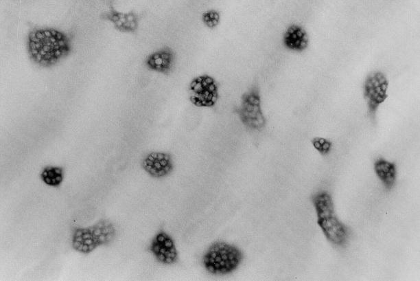
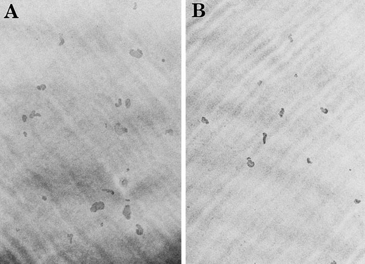
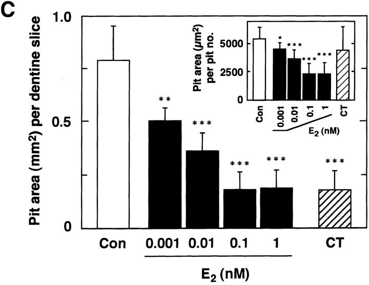
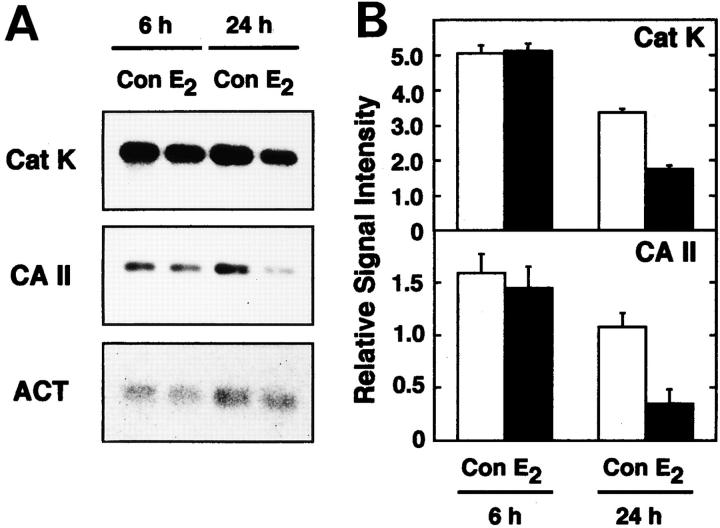
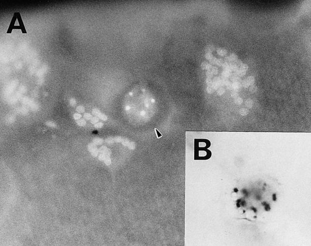
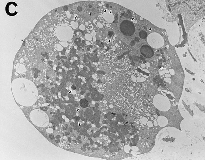
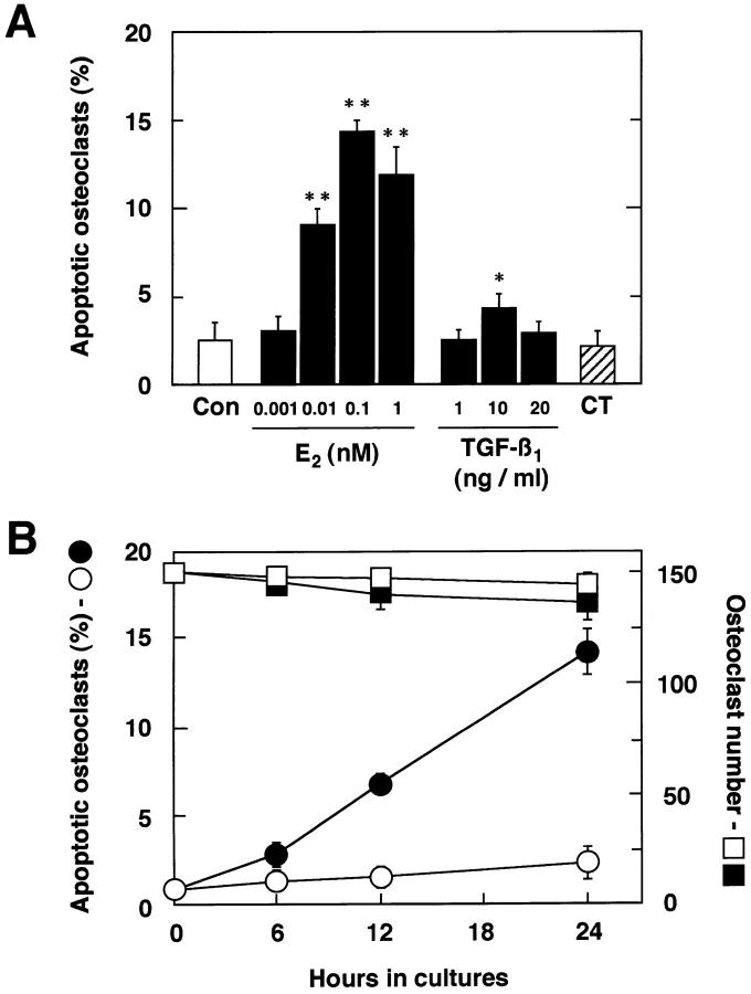
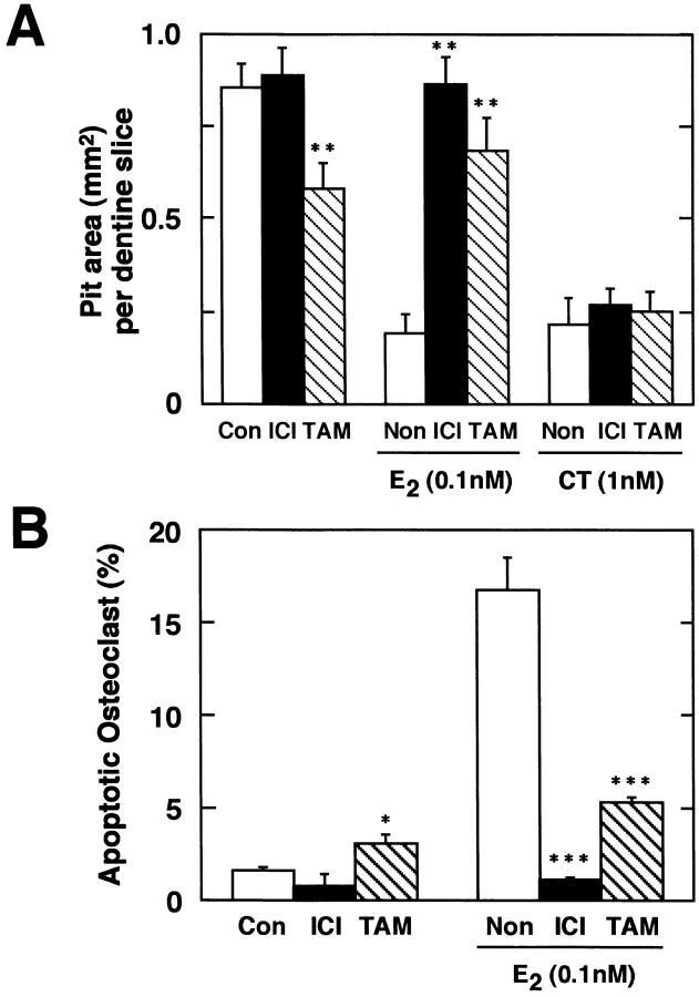
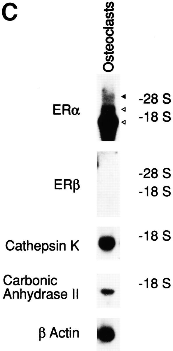
Estrogen deficiency causes bone loss, which can be prevented by estrogen replacement therapy. Using a recently developed technique for isolation of highly purified mammalian osteoclasts, we showed that 17 beta-estradiol (E2) was able to directly inhibit osteoclastic bone resorption. At concentrations effective for inhibiting bone resorption, E2 also directly induced osteoclast apoptosis in a dose- and time-dependent manner. ICI164,384 and tamoxifen, as pure and partial antagonists, respectively, completely or partially blocked the effect of E2 on both inhibition of osteoclastic bone resorption and induction of osteoclast apoptosis. These data suggest that the protective effects of estrogen against postmenopausal osteoporosis are mediated in part by the direct induction of apoptosis of the bone-resorbing osteoclasts by an estrogen receptor- mediated mechanism.


Figures

Figure 1
Figure 1
Purified osteoclasts harvested from collagen gels. 2,000 osteoclasts were plated on a dentine slice and stained for TRAcP activity after a 2-h incubation. (×100).
Figure 2
Figure 2
Estrogen inhibition of bone resorption by purified rabbit osteoclasts. Purified osteoclasts were incubated with medium (A) lacking or (B) containing 0.1 nM E2 for 24 h on dentine slices (×40). After removal of osteoclasts, resorption pits formed by osteoclasts were stained with acid hematoxylin and observed under a light microscope. (C) Inhibitory effects of E2 on osteoclast-mediated bone resorption. Purified osteoclasts were cultured on dentine slices (150 cells/slice) in medium (Con) or in medium containing 0.001–1 nM E2 (E2) or 1 nM salmon calcitonin (CT). Osteoclastic bone resorption activity was measured in terms of pit area formed by purified osteoclasts after 24 h of incubation. Pit area per pit number is also indicated (inset). Values are means ± SD, n = 4. *P <0.05, **P <0.01, ***P <0.005 compared with control groups. Data are representatives of those obtained in three additional independent experiments.
Figure 2
Figure 2
Estrogen inhibition of bone resorption by purified rabbit osteoclasts. Purified osteoclasts were incubated with medium (A) lacking or (B) containing 0.1 nM E2 for 24 h on dentine slices (×40). After removal of osteoclasts, resorption pits formed by osteoclasts were stained with acid hematoxylin and observed under a light microscope. (C) Inhibitory effects of E2 on osteoclast-mediated bone resorption. Purified osteoclasts were cultured on dentine slices (150 cells/slice) in medium (Con) or in medium containing 0.001–1 nM E2 (E2) or 1 nM salmon calcitonin (CT). Osteoclastic bone resorption activity was measured in terms of pit area formed by purified osteoclasts after 24 h of incubation. Pit area per pit number is also indicated (inset). Values are means ± SD, n = 4. *P <0.05, **P <0.01, ***P <0.005 compared with control groups. Data are representatives of those obtained in three additional independent experiments.
Figure 3
Figure 3
Negative regulation of mRNA levels for Cat K and CA II by estrogen. (A) Northern blot analysis. Total RNAs (3 μg) from ∼25,000 purified osteoclasts cultured on dentine slices (150 cells/dentine slices) with (E2) or without 0.1 nM E2 (Con) for 6 or 24 h were used as samples. (B) Relative abundance of Cat K and CA II mRNAs. The relative abundance of Cat K and CA II mRNAs on Northern blotting was evaluated from the values of densitometric scanning. The values shown were normalized with respect to the abundance of β-actin (ACT), and expressed as means ± SD of samples from three other independent experiments.
Figure 4
Figure 4
Estrogen-induced osteoclast apoptosis. Osteoclasts were cultured on dentine slices with 0.1 nM E2 for 24 h. (A) A fluorescence micrograph shows normal and apoptotic osteoclasts (arrowhead) attached to a dentine slice in a 24-h culture. ×250. (B) The TUNEL assay indicates DNA fragmentation in an apoptotic osteoclast. ×250. (C) This micrograph, obtained by electron transmission microscopy, demonstrates the gross morphological changes in an apoptotic osteoclast. Nuclear fragments are indicated by the arrowheads. ×2,000.
Figure 4
Figure 4
Estrogen-induced osteoclast apoptosis. Osteoclasts were cultured on dentine slices with 0.1 nM E2 for 24 h. (A) A fluorescence micrograph shows normal and apoptotic osteoclasts (arrowhead) attached to a dentine slice in a 24-h culture. ×250. (B) The TUNEL assay indicates DNA fragmentation in an apoptotic osteoclast. ×250. (C) This micrograph, obtained by electron transmission microscopy, demonstrates the gross morphological changes in an apoptotic osteoclast. Nuclear fragments are indicated by the arrowheads. ×2,000.
Figure 5
Figure 5
Dose- and time-dependent manner of estrogen-induced osteoclast apoptosis. (A) Purified osteoclasts were cultured on dentine slices (150 cells/slice) for 24 h in medium (Con) or in medium containing 0.001–1 nM E2, 1–20 ng/ml TGF-β1, or 1 nM CT. Apoptotic osteoclasts were quantified under a fluorescence microscope. (B) Time-dependent effects of E2 on osteoclast apoptosis and osteoclast number. Under the same culture conditions, purified osteoclasts were incubated in medium without 0.1 nM E2 (apoptosis: ○; cell number: □) or with 0.1 nM E2 (apoptosis: •, cell number: ▪) for 6, 12, or 24 h. Apoptotic osteoclasts are expressed as a percentage of total number of adherent osteoclasts. Values are means ± SD, n = 4. *P <0.05, **P <0.005 compared with time = 0 groups. Data are representative of those of three additional independent experiments.
Figure 6
Figure 6
Estrogen affects osteoclasts through ER-mediated mechanisms. (A) Antiestrogens recovered E2-inhibited osteoclastic bone resorption. ICI at 1 nM or 1 nM TAM was added to osteoclast cultures in the presence or absence of 0.1 nM E2 or 1 nM salmon CT. 24 h later, osteoclastic bone resorption activity was measured in terms of pit area. (B) Antiestrogens blocked E2-induced osteoclast apoptosis. Under the same culture conditions, purified osteoclasts were incubated in medium with or without 1 nM ICI or 1 nM TAM in the presence or absence of 0.1 nM E2, and osteoclast apoptosis was measured after a 24-h treatment. Apoptotic osteoclasts are expressed as a percentage of total number of adherent osteoclasts. (C) Northern blot analysis of ERα and ERβ mRNA expression in osteoclasts. Total RNA (50 μg) from ∼5 × 10⁵ purified osteoclasts cultured on tissue culture dishes with 0.1 nM E2 for 6 h were used as samples. Values are means ± SD, n = 4. *P <0.05, **P <0.01, ***P <0.005 compared with control or estrogen-treated groups for A and B. Data are representative of those of three additional independent experiments.
Figure 6
Figure 6
Estrogen affects osteoclasts through ER-mediated mechanisms. (A) Antiestrogens recovered E2-inhibited osteoclastic bone resorption. ICI at 1 nM or 1 nM TAM was added to osteoclast cultures in the presence or absence of 0.1 nM E2 or 1 nM salmon CT. 24 h later, osteoclastic bone resorption activity was measured in terms of pit area. (B) Antiestrogens blocked E2-induced osteoclast apoptosis. Under the same culture conditions, purified osteoclasts were incubated in medium with or without 1 nM ICI or 1 nM TAM in the presence or absence of 0.1 nM E2, and osteoclast apoptosis was measured after a 24-h treatment. Apoptotic osteoclasts are expressed as a percentage of total number of adherent osteoclasts. (C) Northern blot analysis of ERα and ERβ mRNA expression in osteoclasts. Total RNA (50 μg) from ∼5 × 10⁵ purified osteoclasts cultured on tissue culture dishes with 0.1 nM E2 for 6 h were used as samples. Values are means ± SD, n = 4. *P <0.05, **P <0.01, ***P <0.005 compared with control or estrogen-treated groups for A and B. Data are representative of those of three additional independent experiments.

References

    1. Lindsay R, Hart DM, Aitken JM, MacDonald EB, Anderson JB, Clarke AC. Long-term prevention of postmenopausal osteoporosis by oestrogen. Evidence for an increased bone mass after delayed onset of oestrogen treatment. Lancet. 1976;1:1038–1041. - PubMed
    1. Ettinger B, Genant HK, Cann CE. Long-term estrogen replacement therapy prevents bone loss and fractures. Ann Intern Med. 1985;102:319–324. - PubMed
    1. Heaney RP, Recker RR, Saville PD. Menopausal changes in bone remodeling. J Lab Clin Med. 1978;92:964–970. - PubMed
    1. Selby PL, Peacock M, Barkworth SA, Brown WB, Taylor GA. Early effects of ethinyloestradiol and norethisterone treatment in post-menopausal women on bone resorption and calcium regulating hormones. Clin Sci (Lond) 1985;69:265–271. - PubMed
    1. Vaes G. Cellular biology and biochemical mechanism of bone resorption. A review of recent developments on the formation, activation, and mode of action of osteoclasts. Clin Orthop Relat Res. 1988;231:239–271. - PubMed