. 1997 Aug 18;186(4):517-27.
doi: 10.1084/jem.186.4.517.

Endothelial-dependent mechanisms regulate leukocyte transmigration: a process involving the proteasome and disruption of the vascular endothelial-cadherin complex at endothelial cell-to-cell junctions

Affiliations

Endothelial-dependent mechanisms regulate leukocyte transmigration: a process involving the proteasome and disruption of the vascular endothelial-cadherin complex at endothelial cell-to-cell junctions

J R Allport et al. J Exp Med. .

Abstract

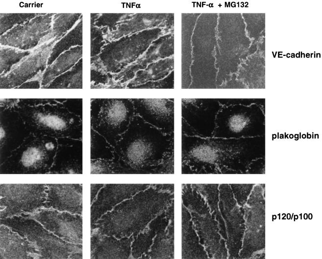
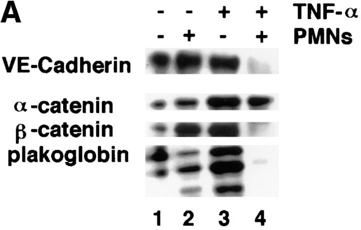
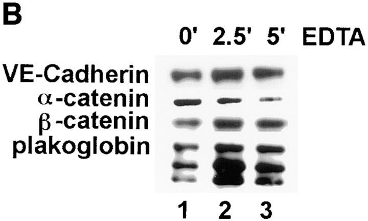
Although several adhesion molecules expressed on leukocytes (beta1 and beta2 integrins, platelet endothelial cell adhesion molecule 1 [PECAM-1], and CD47) and on endothelium (intercellular adhesion molecule 1, PECAM-1) have been implicated in leukocyte transendothelial migration, less is known about the role of endothelial lateral junctions during this process. We have shown previously (Read, M.A., A.S. Neish, F.W. Luscinskas, V.J. Palambella, T. Maniatis, and T. Collins. 1995. Immunity. 2:493-506) that inhibitors of the proteasome reduce lymphocyte and neutrophil adhesion and transmigration across TNF-alpha-activated human umbilical vein endothelial cell (EC) monolayers in an in vitro flow model. The current study examined EC lateral junction proteins, principally the vascular endothelial (VE)-cadherin complex and the effects of proteasome inhibitors (MG132 and lactacystin) on lateral junctions during leukocyte adhesion, to gain a better understanding of the role of EC junctions in leukocyte transmigration. Both biochemical and indirect immunofluorescence analyses of the adherens junction zone of EC monolayers revealed that neutrophil adhesion, not transmigration, induced disruption of the VE-cadherin complex and loss of its lateral junction localization. In contrast, PECAM-1, which is located at lateral junctions and is implicated in neutrophil transmigration, was not altered. These findings identify new and interrelated endothelial-dependent mechanisms for leukocyte transmigration that involve alterations in lateral junction structure and a proteasome-dependent event(s).


Figures

Figure 1
Figure 1
Proteasome inhibitors MG132 and lactacystin prevent neutrophil migration under flow at 1.8 dynes/cm2. Confluent EC monolayers were incubated with TNF-α for 4 h before addition of proteasome inhibitors (MG, 20 μM MG132; LAC, 20 μM lactacystin) or DMSO carrier (Cont.) for 1 (left) or 2 h (right), and neutrophil adhesion and transmigration were assessed as detailed in Materials and Methods. *P <0.05.
Figure 2
Figure 2
The endothelial VE–cadherin complex is not altered by incubation with proteasomal inhibitors with or without TNF-α treatment. Confluent EC monolayers were treated with inhibitors before or after addition of TNF-α, washed twice, and extracted in lysis buffer 1 (Materials and Methods). The VE–cadherin complex was immunoprecipitated using anti–VE–cadherin mAb, separated by SDS-PAGE, and subjected to immunoblotting using mAbs that recognize α- and β-catenins, plakoglobin, and p120. The results with anti-p120 mAb were variable and p120 was not always detected, even though the other components shown were consistent and reproducible. The x-axis labels are: C, 0.02% DMSO; T, 25 ng/ml of TNF-α for 4 h; MG, MG132 (20 μM) for 4 h; ALLM, ALLM (20 μM) treatment for 4 h.
Figure 3
Figure 3
Colocalization of VE–cadherin, plakoglobin, and p120/p100 to lateral cell-to-cell junctions in confluent EC monolayers is not altered by TNF-α alone or TNF-α plus MG132. Confluent cultured EC were treated with carrier (0.02% DMSO), TNF-α (4 h), or TNF-α (4 h) followed by 2 h of incubation with MG132 (20 μM), washed, and fixed on ice with cold methanol for 5 min. Complex components were detected using specific mAb as detailed in Materials and Methods.
Figure 4
Figure 4
Effect of neutrophil adhesion on the endothelial VE–cadherin complex. (A) Confluent EC monolayers were incubated with media alone or media containing TNF-α for 6 h and washed. Neutrophils (107) in DPBS-HSA or DPBS-HSA alone were added for 10 min at 37°C under static conditions. Nonadherent neutrophils were removed by washing and the monolayers were extracted on ice for 30 min. Proteins were immunoprecipitated with anti–VE–cadherin mAb as detailed in Materials and Methods and detected by immunoblotting with specific mAb. (B) Confluent EC monolayers were treated with media alone or media containing TNF-α for 4 h, washed twice, and 107 neutrophils were added. After incubation for 0, 3, or 10 min at 37°C, nonadherent neutrophils were removed, and monolayers were extracted with lysis buffer 1 containing 0.5% SDS. Proteins were immunoprecipitated using specific mAb directed against each individual component and subsequently detected by immunoblotting with a specific mAb. The Mr for molecular mass standards are shown on the left margin and 0, 3, and 10 refer to the time period of neutrophil–EC coincubation at 37°C. (C) Media alone (lane 1), resting neutrophils (107 cells, lane 2), neutrophil membranes representing 107 cells (lane 3), or conditioned media from coincubations of neutrophils (107 cells) with TNF-α–activated HUVEC (lane 4) were added to control (lane 1) or TNF-α–activated HUVEC monolayers (lanes 2–4) and incubated at 37°C for 10 min. HUVEC were washed twice and extracted with ice-cold lysis buffer 1 (without 0.5% SDS). Proteins were immunoprecipitated with anti–VE–cadherin mAb as detailed in Materials and Methods, and detected by immunoblotting with specific mAb. (D) Neutrophil membranes representing 3 × 10⁶ cells were incubated with 4 h TNF-α–activated EC in wells of chamber slides for 10 min at 37°C. The monolayers were washed twice and fixed for 5 min at 4°C with ice-cold methanol. Junctional proteins were detected by indirect immunofluorescence.
Figure 4
Figure 4
Effect of neutrophil adhesion on the endothelial VE–cadherin complex. (A) Confluent EC monolayers were incubated with media alone or media containing TNF-α for 6 h and washed. Neutrophils (107) in DPBS-HSA or DPBS-HSA alone were added for 10 min at 37°C under static conditions. Nonadherent neutrophils were removed by washing and the monolayers were extracted on ice for 30 min. Proteins were immunoprecipitated with anti–VE–cadherin mAb as detailed in Materials and Methods and detected by immunoblotting with specific mAb. (B) Confluent EC monolayers were treated with media alone or media containing TNF-α for 4 h, washed twice, and 107 neutrophils were added. After incubation for 0, 3, or 10 min at 37°C, nonadherent neutrophils were removed, and monolayers were extracted with lysis buffer 1 containing 0.5% SDS. Proteins were immunoprecipitated using specific mAb directed against each individual component and subsequently detected by immunoblotting with a specific mAb. The Mr for molecular mass standards are shown on the left margin and 0, 3, and 10 refer to the time period of neutrophil–EC coincubation at 37°C. (C) Media alone (lane 1), resting neutrophils (107 cells, lane 2), neutrophil membranes representing 107 cells (lane 3), or conditioned media from coincubations of neutrophils (107 cells) with TNF-α–activated HUVEC (lane 4) were added to control (lane 1) or TNF-α–activated HUVEC monolayers (lanes 2–4) and incubated at 37°C for 10 min. HUVEC were washed twice and extracted with ice-cold lysis buffer 1 (without 0.5% SDS). Proteins were immunoprecipitated with anti–VE–cadherin mAb as detailed in Materials and Methods, and detected by immunoblotting with specific mAb. (D) Neutrophil membranes representing 3 × 10⁶ cells were incubated with 4 h TNF-α–activated EC in wells of chamber slides for 10 min at 37°C. The monolayers were washed twice and fixed for 5 min at 4°C with ice-cold methanol. Junctional proteins were detected by indirect immunofluorescence.
Figure 5
Figure 5
Chelation of extracellular Ca2+ by EDTA releases the VE–cadherin complex from the lateral junctions, but does not trigger degradation. (A) HUVEC monolayers were washed and incubated for 2.5 min with 3 mM EDTA in HBSS, and then fixed for 5 min in ice-cold methanol. VE–cadherin or PECAM-1 was detected by indirect immunofluorescence as detailed in Materials and Methods, and the images were captured using a cooled charged-coupled device camera. (B) Immunoprecipitation and blotting studies show that incubation with EDTA does not induce cleavage of VE–cadherin. Confluent EC monolayers were incubated with TNF-α for 4 h, and then washed twice with DPBS and incubated in either HBSS (lane 1) or HBSS medium containing 3 mM EDTA for 2.5 (lane 2) or 5 min (lane 3) at 37°C. VE–cadherin was immunoprecipitated, resolved by SDS-PAGE, and subjected to blotting as detailed in Fig. 3 legend.
Figure 5
Figure 5
Chelation of extracellular Ca2+ by EDTA releases the VE–cadherin complex from the lateral junctions, but does not trigger degradation. (A) HUVEC monolayers were washed and incubated for 2.5 min with 3 mM EDTA in HBSS, and then fixed for 5 min in ice-cold methanol. VE–cadherin or PECAM-1 was detected by indirect immunofluorescence as detailed in Materials and Methods, and the images were captured using a cooled charged-coupled device camera. (B) Immunoprecipitation and blotting studies show that incubation with EDTA does not induce cleavage of VE–cadherin. Confluent EC monolayers were incubated with TNF-α for 4 h, and then washed twice with DPBS and incubated in either HBSS (lane 1) or HBSS medium containing 3 mM EDTA for 2.5 (lane 2) or 5 min (lane 3) at 37°C. VE–cadherin was immunoprecipitated, resolved by SDS-PAGE, and subjected to blotting as detailed in Fig. 3 legend.
Figure 6
Figure 6
Photographs show VE–cadherin, β-catenin, p120/ p100, plakoglobin, and PECAM-1 staining patterns. Indirect immunofluorescence microscopy shows that TNF-α–activated EC monolayers express abundant levels of VE–cadherin, β-catenin, p120/ p100, plakoglobin, and PECAM-1 at lateral junctions before neutrophil adhesion (left). After 10 min of neutrophil adhesion, the lateral staining of VE–cadherin, β-catenin, p120/p100, and plakoglobin is lost, whereas PECAM-1 is still abundantly expressed at lateral junctions (right). Adherent neutrophils are identified (right, arrows), and intact VE–cadherin junctional staining (top asterisks). Original magnification was 320, except VE–cadherin which was 640.
Figure 7
Figure 7
Proteasome inhibitors do not prevent neutrophil-induced disruption of the VE–cadherin complex. Neutrophils (107 in 5 ml) were incubated with EC monolayers that were treated with TNF-α (4 h, 25 ng/ml) followed by 2 h of incubation with MG132 (M), ALLM (A), or lactacystin (L), or 0.02% DMSO (−). After 10 min at 37°C, EC monolayers were extracted with lysis buffer 1 and processed for detection of the VE–cadherin complex.

References

    1. Bevilacqua MP, Pober JS, Wheeler ME, Cotran RS, Gimbrone MA., Jr Interleukin-1 acts on cultured human vascular endothelium to increase the adhesion of polymorphonuclear leukocytes, monocytes, and related leukocyte cell lines. J Clin Invest. 1985;76:2003–2011. - PMC - PubMed
    1. Schleimer RP, Rutledge RK. Cultured human vascular endothelial cells acquire adhesiveness for leukocytes following stimulation with interleukin-1, endotoxin and tumor-promoting phorbol-esters. J Immunol. 1986;136:649–656. - PubMed
    1. Gamble JR, Harlan JM, Klebanoff SJ, Lopez AF, Vadas MA. Stimulation of the adherence of neutrophils to umbilical vein endothelium by recombinant tumor necrosis factor. Proc Natl Acad Sci USA. 1985;82:8667–8670. - PMC - PubMed
    1. Read MA, Neish AS, Luscinskas FW, Palombella VJ, Maniatis T, Collins T. The proteasome pathway is required for cytokine-induced endothelial–leukocyte adhesion molecule expression. Immunity. 1995;2:493–506. - PubMed
    1. Dejana E, Corada M, Lampugnani G. Endothelial cell-to-cell junctions. FASEB J. 1995;9:910–918. - PubMed

Publication types