. 1997 Aug 18;186(4):589-600.
doi: 10.1084/jem.186.4.589.

Vascular adhesion protein 1 (VAP-1) mediates lymphocyte subtype-specific, selectin-independent recognition of vascular endothelium in human lymph nodes

Affiliations

Vascular adhesion protein 1 (VAP-1) mediates lymphocyte subtype-specific, selectin-independent recognition of vascular endothelium in human lymph nodes

M Salmi et al. J Exp Med. .

Abstract

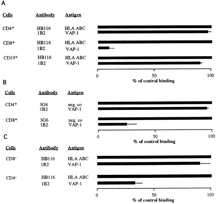
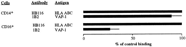
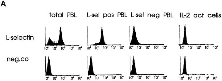
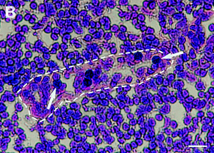
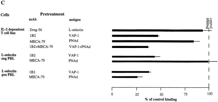
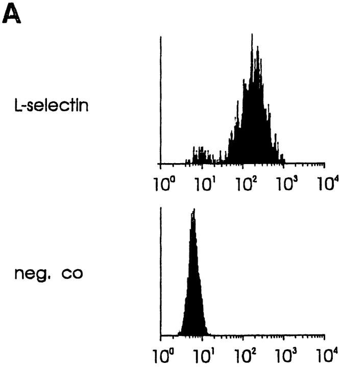
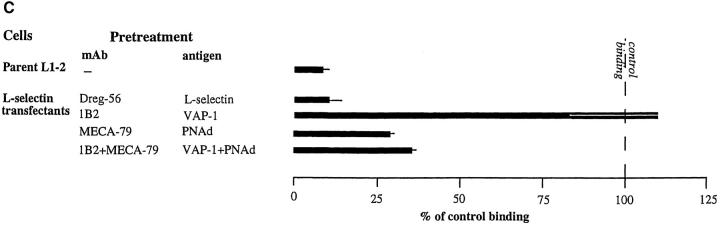
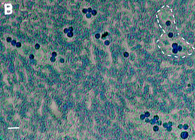
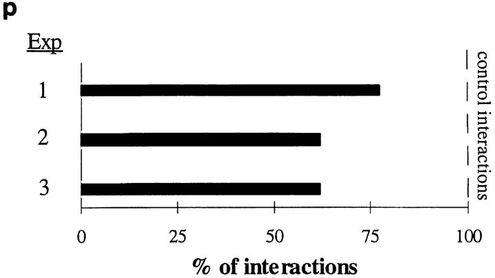
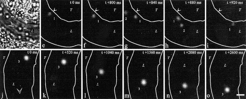
Interactions between lymphocyte surface receptors and their ligands on vascular endothelial cells regulate the exit of lymphocytes from the circulation. Distinct subsets of mononuclear cells bind to high endothelial venules (HEVs) in different lymphoid organs to a different extent, but the molecular mechanisms behind this selectivity have remained poorly characterized. Here we show that vascular adhesion protein-1 (VAP-1) mediates subtype-specific binding of CD8-positive T cells and natural killer cells to human endothelium. VAP-1-dependent, oligosaccharide-dependent peripheral lymph node (PLN) HEV adhesion under shear was independent of L-selectin, P-selectin glycoprotein ligand 1, and alpha4 integrins, the known lymphocyte receptors involved in the initial recognition of endothelial cells. PLN HEV adhesion was also critically dependent on peripheral lymph node vascular addressins (PNAds), but lymphocyte L-selectin was absolutely required for PNAd binding. Most lymphocytes relied on both PNAd and VAP-1 in HEV binding. The overlapping function of L-selectin ligands and VAP-1 in PLN introduces a new control point into the lymphocyte extravasation process. Finally, intravital microscopy revealed that VAP-1 is involved in initial interactions between human lymphocytes and endothelial cells in inflamed rabbit mesenterial venules in vivo. In conclusion, VAP-1 is a novel contact-initiating ligand that discriminates between different subpopulations of mononuclear cells and is an appealing target for selective modulation of adhesion of CD8- and CD16-positive effector cells.


Figures

Figure 1
Figure 1
VAP-1 selectively mediates HEV adhesion of CD8-positive lymphocytes. (A) CD4-, CD8-, and CD19-positive PBL were isolated with MACS, and their adherence to PLN HEVs pretreated with a control or an anti–VAP-1 mAb was tested as detailed in Materials and Methods. (B) PBL were labeled with FITC-conjugated anti-CD4 or -CD8 mAbs, and the number of labeled cells adherent to PLN HEVs in the presence of control and anti–VAP-1 mAbs was determined. (C) PBL were subjected to negative selection (removal of CD8-positive cells yielding a CD8-negative population for analysis (CD8 ), or removal of CD4-positive cells yielding a CD4-negative population (CD4 ) and the effect of anti–VAP-1 and control mAb pretreatments on HEV adhesion was determined.
Figure 2
Figure 2
NK, but not monocytes, use VAP-1 for endothelial binding. CD14- and CD16-positive PBL were isolated with MACS and their binding to tonsil HEVs pretreated with a control or anti–VAP-1 mAb was tested.
Figure 3
Figure 3
VAP-1 mediates binding of both L-selectin–positive and –negative lymphocytes to PLN HEV. (A) Expression of L-selectin on different lymphocyte populations. Total PBL, immunomagnetically separated L-selectin–positive and –negative subpopulations of PBL, and IL-2–activated T cells were stained for L-selectin expression and analyzed using FACScan®. (B) L-selectin–negative T cells specifically recognize PLN HEVs. Five immunoblasts (black round cells, two pointed out by arrows) binding to a HEV (lilac basement membrane outlined by a dashed line) are shown in the micrograph. Bar, 20 μm. (C) Binding of these different lymphocytes to PLN HEVs after pretreatments with the indicated mAbs was analyzed.
Figure 3
Figure 3
VAP-1 mediates binding of both L-selectin–positive and –negative lymphocytes to PLN HEV. (A) Expression of L-selectin on different lymphocyte populations. Total PBL, immunomagnetically separated L-selectin–positive and –negative subpopulations of PBL, and IL-2–activated T cells were stained for L-selectin expression and analyzed using FACScan®. (B) L-selectin–negative T cells specifically recognize PLN HEVs. Five immunoblasts (black round cells, two pointed out by arrows) binding to a HEV (lilac basement membrane outlined by a dashed line) are shown in the micrograph. Bar, 20 μm. (C) Binding of these different lymphocytes to PLN HEVs after pretreatments with the indicated mAbs was analyzed.
Figure 4
Figure 4
VAP-1 does not recognize L-selectin. (A) Expression of human L-selectin on mouse L1-2 L-selectin transfectants. (B) L1-2 L-selectin transfectants (dark blue spots) efficiently bind to human PLN HEVs (seven different-sized HEV, one outlined by a dashed line, can be seen as light blue structures). (C) L1-2 L-selectin transfectant binding to HEV in human PLNs is independent of VAP-1. Transfectants were pretreated with Dreg-56 and PLN tissue with 1B2 and/or MECA-79, and the HEV adhesion was determined. (D) L-selectin chimera binds PNAd, but not VAP-1. The Ig-chimeras were used to deplete tonsil lysate and the nonprecipitated molecules were analyzed using immunoblotting and the mAbs indicated.
Figure 4
Figure 4
VAP-1 does not recognize L-selectin. (A) Expression of human L-selectin on mouse L1-2 L-selectin transfectants. (B) L1-2 L-selectin transfectants (dark blue spots) efficiently bind to human PLN HEVs (seven different-sized HEV, one outlined by a dashed line, can be seen as light blue structures). (C) L1-2 L-selectin transfectant binding to HEV in human PLNs is independent of VAP-1. Transfectants were pretreated with Dreg-56 and PLN tissue with 1B2 and/or MECA-79, and the HEV adhesion was determined. (D) L-selectin chimera binds PNAd, but not VAP-1. The Ig-chimeras were used to deplete tonsil lysate and the nonprecipitated molecules were analyzed using immunoblotting and the mAbs indicated.
Figure 5
Figure 5
VAP-1 binds to a novel contact initiating lymphocyte counterreceptor. PBL were treated with the indicated mAbs (HP2/1 and PL1) against lymphocyte adhesion receptors or with O-sialoglycoprotease (O-sgp), and their binding to the target tissue treated with anti–VAP-1 or control mAbs was evaluated. Binding of PBL treated with anti–α4 integrin mAb was tested to PLN HEVs, whereas binding of PBL pretreated with anti– PSGL-1 mAb and O-sgp was evaluated using tonsil as a target tissue.
Figure 6
Figure 6
Most PBL use both PNAd and VAP-1 in PLN HEV binding. PBL were treated with anti–L-selectin mAb and PLNs with mAbs against different endothelial adhesion molecules alone or in combination, and the HEV adherence was determined.
Figure 7
Figure 7
CD8-positive PBL use both L-selectin and VAP-1 to adhere to PLN HEVs. Positively selected CD8-positive cells (CD8 +) were treated with anti– L-selectin and control mAb, PLN tissue section was pretreated with anti–VAP-1 and control mAb, and the HEV binding was determined.
Figure 8
Figure 8
VAP-1 mediates initial interactions between human lymphocytes and inflamed vascular endothelium in vivo. (a) An anti–human VAP-1 mAb 5B11 recognizes VAP-1 in HEVs in frozen sections of rabbit mesenterial lymph node. (b) Negative control staining with mAb 4D7. An HEV is pointed out by an arrow. (c) Rabbit VAP-1 is expressed on the luminal surface of inflamed mesenterial vessel. After intravenous injection of mAb 5B11, the in vivo–bound mAb was immunohistochemically detected in the mesenterial section with a second-stage peroxidase-conjugated anti–mouse Ig. (d–o) Intravital microscopy was used to study the effect of a control mAb 4D7 and an anti–VAP-1 mAb 5B11 on binding of fluorescently labeled human tonsillar lymphocytes with inflamed mesenterial vessels in rabbits as detailed in Materials and Methods. (d) A micrograph of one segment of the vessel under study. L, lumen of the vessel; T, connective tissue of the mesenterium; white triangles, the walls of the vessel; white arrows, rolling rabbit granulocytes. (e–i) A tethering lymphocyte. The same segment as in d viewed under stroboscopic epiillumination after injecting fluorescently labeled human cells. A cell (1) docks to the vessel wall in e, moves less than 5 μm/s during the first 800 ms (e–f), speeds up in g, and detaches in h–i. A freely flowing cell (2; velocity 1,110 μm/s) is also seen. Arrows mark a reference point at the vessel wall. (j–o) A rolling lymphocyte. In another segment of a venule, a lymphocyte (3) that rolls along the bottom of the vessel for >2.5 s is seen (average velocity ∼24 μm/s). Blood flow (open arrowhead) is from left to right in d–i and from top to bottom in j–o. White lines, vessel walls. Time codes in the upper right corners indicate the time elapsed (in ms) from the first frame of the series. Bar, 10 μm. (p) Anti–VAP-1 mAb 5B11 reduces interactions between human lymphocytes and rabbit vessel wall when analyzed by intravital microscopy in three independent experiments. Number of control interactions was arbitrarily set at 100%.
Figure 8
Figure 8
VAP-1 mediates initial interactions between human lymphocytes and inflamed vascular endothelium in vivo. (a) An anti–human VAP-1 mAb 5B11 recognizes VAP-1 in HEVs in frozen sections of rabbit mesenterial lymph node. (b) Negative control staining with mAb 4D7. An HEV is pointed out by an arrow. (c) Rabbit VAP-1 is expressed on the luminal surface of inflamed mesenterial vessel. After intravenous injection of mAb 5B11, the in vivo–bound mAb was immunohistochemically detected in the mesenterial section with a second-stage peroxidase-conjugated anti–mouse Ig. (d–o) Intravital microscopy was used to study the effect of a control mAb 4D7 and an anti–VAP-1 mAb 5B11 on binding of fluorescently labeled human tonsillar lymphocytes with inflamed mesenterial vessels in rabbits as detailed in Materials and Methods. (d) A micrograph of one segment of the vessel under study. L, lumen of the vessel; T, connective tissue of the mesenterium; white triangles, the walls of the vessel; white arrows, rolling rabbit granulocytes. (e–i) A tethering lymphocyte. The same segment as in d viewed under stroboscopic epiillumination after injecting fluorescently labeled human cells. A cell (1) docks to the vessel wall in e, moves less than 5 μm/s during the first 800 ms (e–f), speeds up in g, and detaches in h–i. A freely flowing cell (2; velocity 1,110 μm/s) is also seen. Arrows mark a reference point at the vessel wall. (j–o) A rolling lymphocyte. In another segment of a venule, a lymphocyte (3) that rolls along the bottom of the vessel for >2.5 s is seen (average velocity ∼24 μm/s). Blood flow (open arrowhead) is from left to right in d–i and from top to bottom in j–o. White lines, vessel walls. Time codes in the upper right corners indicate the time elapsed (in ms) from the first frame of the series. Bar, 10 μm. (p) Anti–VAP-1 mAb 5B11 reduces interactions between human lymphocytes and rabbit vessel wall when analyzed by intravital microscopy in three independent experiments. Number of control interactions was arbitrarily set at 100%.

References

    1. Gowans JL, Knight EJ. The route of re-circulation of lymphocytes in rat. Proc R Soc Lond Ser B. 1964;159:257–282. - PubMed
    1. Stevens SK, Weissman IL, Butcher EC. Differences in the migration of B and T lymphocytes: organ-selective localization in vivoand the role of lymphocyte– endothelial cell recognition. J Immunol. 1982;128:844–851. - PubMed
    1. Fossum S, Smith ME, Ford WL. The recirculation of T and B lymphocytes in the athymic, nude rat. Scand J Immunol. 1983;17:551–557. - PubMed
    1. van Dinther-Janssen ACHM, van Maarsseveen ACMT, DeGroot J, Scheper RJ. Comparative migration of T- and B-lymphocyte subpopulations into skin inflammatory sites. Immunology. 1983;48:519–527. - PMC - PubMed
    1. Kraal G, Weissman IL, Butcher EC. Differences in in vivo distribution and homing of T cell subsets to mucosal vs.non-mucosal lymphoid organs. J Immunol. 1983;130:1097–1102. - PubMed

Publication types