. 1997 Aug 18;186(4):601-12.
doi: 10.1084/jem.186.4.601.

Kinetics of eotaxin generation and its relationship to eosinophil accumulation in allergic airways disease: analysis in a guinea pig model in vivo

Affiliations

Kinetics of eotaxin generation and its relationship to eosinophil accumulation in allergic airways disease: analysis in a guinea pig model in vivo

A A Humbles et al. J Exp Med. .

Abstract

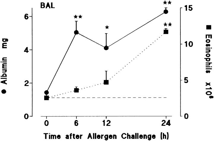
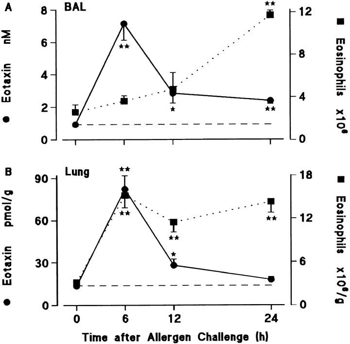
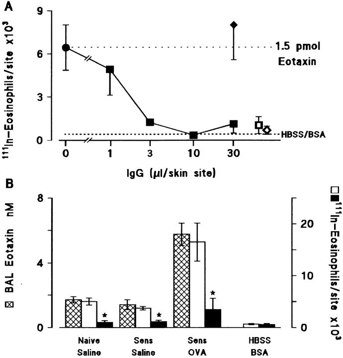
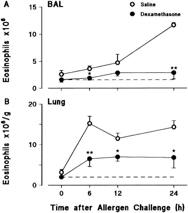
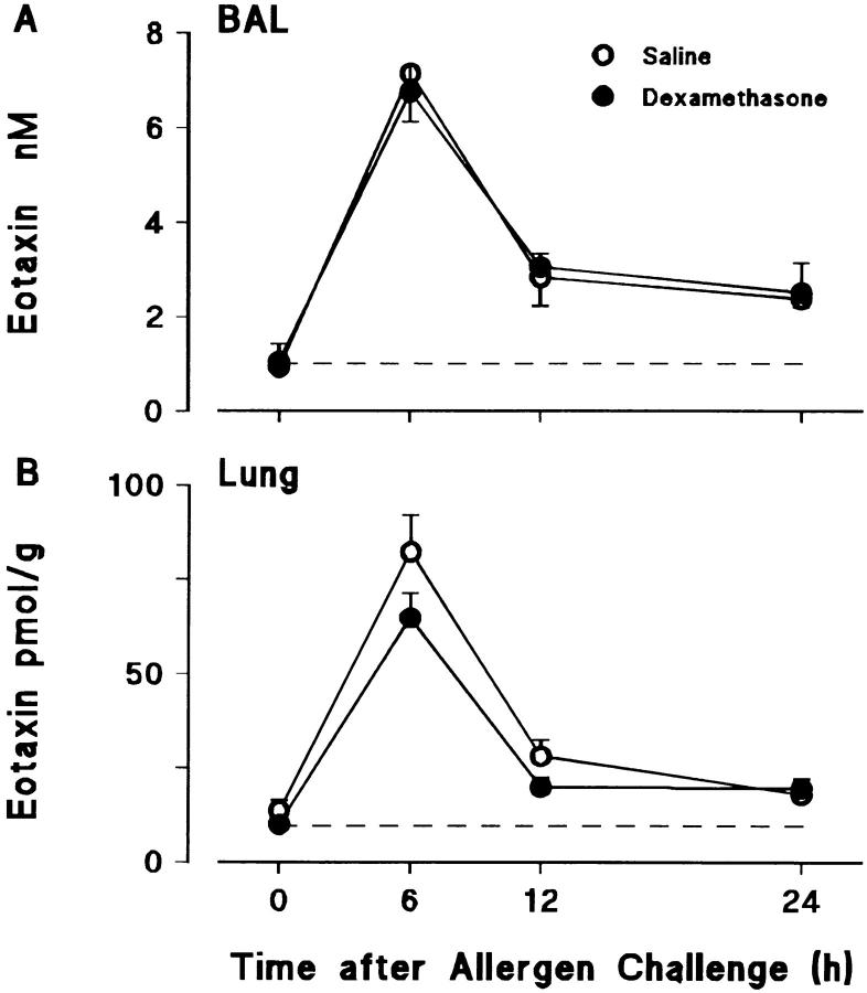
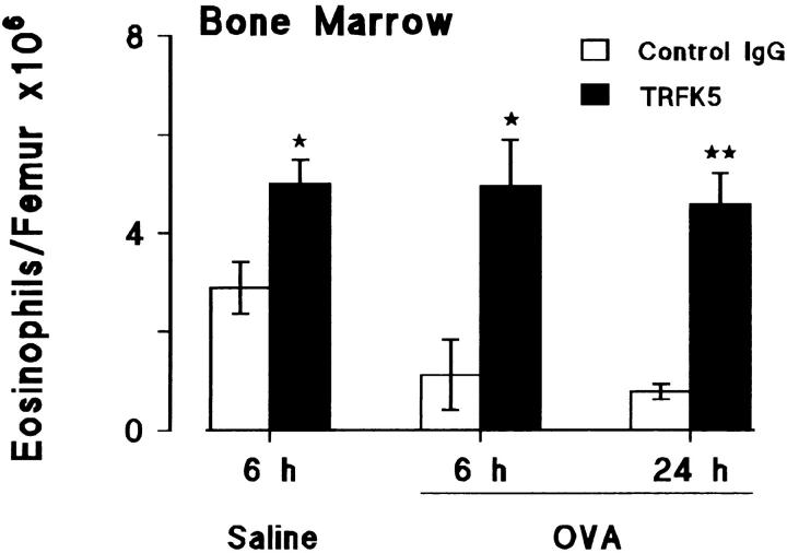
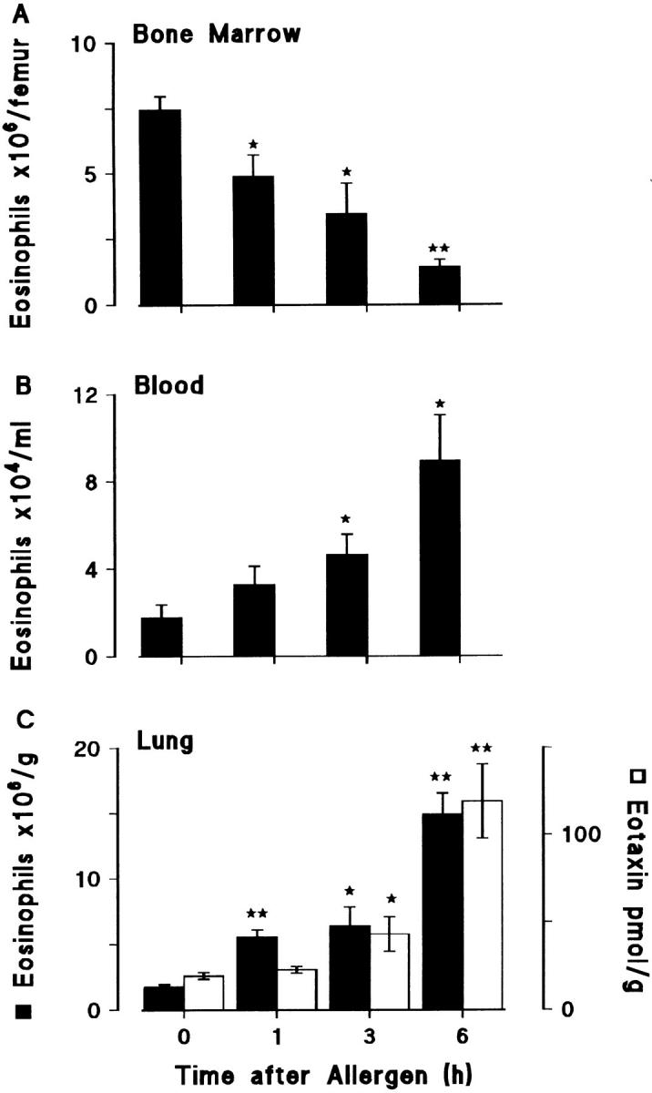
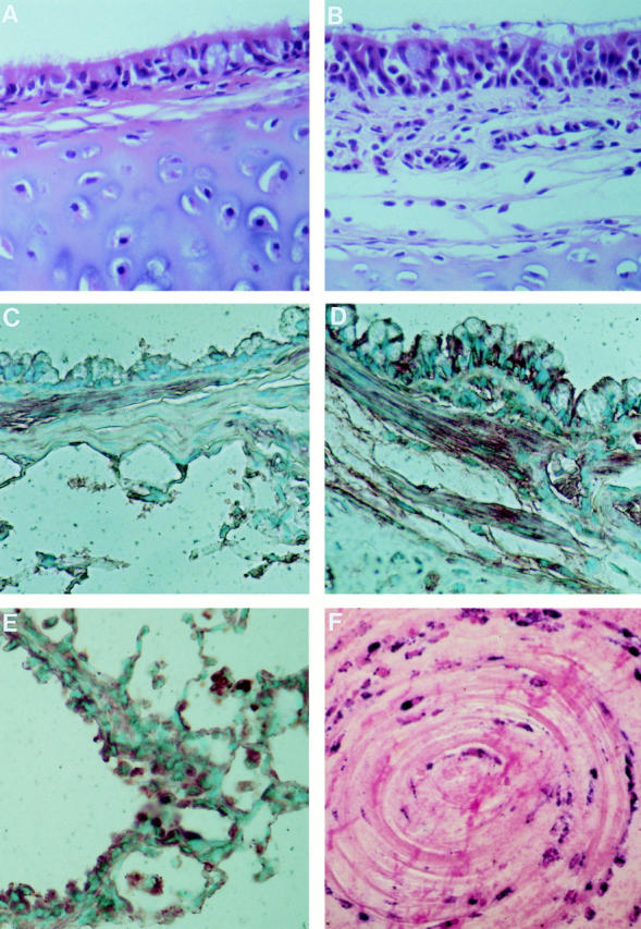
Challenge of the airways of sensitized guinea pigs with aerosolized ovalbumin resulted in an early phase of microvascular protein leakage and a delayed phase of eosinophil accumulation in the airway lumen, as measured using bronchoalveolar lavage (BAL). Immunoreactive eotaxin levels rose in airway tissue and BAL fluid to a peak at 6 h falling to low levels by 12 h. Eosinophil numbers in the tissue correlated with eotaxin levels until 6 h but eosinophils persisted until the last measurement time point at 24 h. In contrast, few eosinophils appeared in BAL over the first 12 h, major trafficking through the airway epithelium occurring at 12-24 h when eotaxin levels were low. Constitutive eotaxin was present in BAL fluid. Both constitutive and allergen-induced eosinophil chemoattractant activity in BAL fluid was neutralized by an antibody to eotaxin. Allergen-induced eotaxin appeared to be mainly in airway epithelium and macrophages, as detected by immunostaining. Allergen challenge of the lung resulted in a rapid release of bone marrow eosinophils into the blood. An antibody to IL-5 suppressed bone marrow eosinophil release and lung eosinophilia, without affecting lung eotaxin levels. Thus, IL-5 and eotaxin appear to cooperate in mediating a rapid transfer of eosinophils from the bone marrow to the lung in response to allergen challenge.


Figures

Figure 1
Figure 1
Time course of albumin extravasation (•, solid line) and eosinophil accumulation (▪, dotted line) in BAL after allergen challenge. Sensitized guinea pigs were exposed to aerosolized ovalbumin and killed at various times after challenge. Results are presented as a mean±SEM of 5–11 animals/time point. 0 h represents sensitized/nonchallenged animals (n = 5). Significant differences from 0 h are indicated as *P <0.02 and **P <0.005. Basal eosinophil numbers in BAL are shown by the dashed line.
Figure 2
Figure 2
Time course of eotaxin generation (•, solid line) and eosinophil accumulation (▪, dotted line) in (A) BAL and (B) lung tissue of sensitized guinea pigs after allergen challenge. Results are presented as a mean±SEM (n = 5–9 animals/time point). 0 h represents sensitized/ nonchallenged animals (n = 5). Significant differences from 0 h are indicated as *P <0.05 and **P <0.005. Constitutive levels of eotaxin in BAL fluid and lung tissue are shown by the dashed lines.
Figure 3
Figure 3
(A) Inhibition of eotaxin-induced 111In-eosinophil accumulation by antieotaxin antibody. Purified eotaxin (1.5 pmol) was coinjected with antieotaxin IgG (▪, 1–30 μl), control IgG (♦, 30 μl) or without IgG (•, upper dotted line) into the skin of naive guinea pigs that had received an i.v. injection of 111In-eosinophils. Results are presented as a mean±SEM of 111In-eosinophils/skin site after 4 h (n = 4–6 bioassay animals). Responses to the intradermal vehicle (HBSS/0.25% BSA) alone are shown by the lower dotted line. Antieotaxin (□, 30 μl) or control IgG (⋄, 30 μl) added to the HBSS/BSA did not induce significant 111In-eosinophil accumulation. (B) Inhibition of 111In-eosinophil chemoattractant activity in 6 h BAL fluid by antieotaxin antibody. BAL fluid obtained 6 h after exposure of naive or sensitized guinea pigs to aerosolized saline or ovalbumin was concentrated by C18 SepPak chromatography (see Materials and Methods) and coinjected with 50 μl control IgG (open bars) or antieotaxin IgG (solid bars) into the skin of naive guinea pigs which had received an i.v. injection of 111In-eosinophils. Responses to HBSS/BSA + control IgG or antieotaxin antibodies were also determined. Results are presented as a mean±SEM (n = 4 bioassay animals) and significant differences between control IgG and antieotaxin IgG treated samples are indicated as *P <0.01. Eotaxin concentrations in unconcentrated BAL fluid (cross hatched bars) are also shown in B. Eotaxin in lung tissue was 28.6 ± 2.5, 24.9 ± 2.1, and 89.6 ± 3.0 pmol/g (mean±SEM, n = 5) in the naive/saline, sensitized/saline, and sensitized/ovalbumin groups, respectively. When these results are expressed as total eotaxin per guinea pig lung, there was approximately five to six times more eotaxin in lung tissue (114 ± 8, 115 ± 16, and 379 ± 14 pmol) than in BAL fluid (23 ± 3, 18 ± 4, and 82 ± 10 pmol, respectively).
Figure 4
Figure 4
Effect of dexamethasone on allergen-induced eosinophil accumulation in (A) BAL and (B) lung tissue. Sensitized guinea pigs were injected i.p. with dexamethasone (40 mg/kg) or saline at 24 and 1 h before allergen challenge. 0 h represents sensitized/nonchallenged animals (n = 5). Results are presented as mean±SEM (n = 5–11 allergen-challenged animals/time point) and significant differences between dexamethasone- and saline-treated groups are indicated as *P <0.05 and **P <0.005. Basal eosinophil numbers in BAL and lung tissue are shown by the dashed lines.
Figure 5
Figure 5
Effect of dexamethasone on allergen-induced eotaxin generation in (A) BAL fluid and (B) lung tissue. Sensitized guinea pigs were injected i.p. with dexamethasone (40 mg/kg) or saline at 24 and 1 h before allergen challenge. 0 h represents sensitized/nonchallenged animals (n = 5). Results are presented as mean±SEM (n = 5–11 allergen-challenged animals/time point). Constitutive levels of eotaxin in BAL and lung tissue are shown by the dashed lines.
Figure 6
Figure 6
Effect of an anti–IL-5 antibody, TRFK5, on eosinophil numbers in (A) BAL and (B) lung tissue, and eotaxin levels in (C) BAL fluid and (D) lung tissue after allergen challenge. Sensitized guinea pigs were injected i.v. with control rat IgG or TRFK5 (0.3 mg/kg) 30 min before aerosol exposure to saline or allergen. Results are presented as mean±SEM (n = 5–6 animals/group) and significant differences between control IgG and TRFK5-treated groups are indicated as *P <0.05 and **P <0.005.
Figure 7
Figure 7
Effect of an anti–IL-5 antibody, TRFK5, on eosinophil numbers in bone marrow after allergen challenge. Sensitized guinea pigs were injected i.v. with control rat IgG or TRFK5 (0.3 mg/kg) 30 min before aerosol exposure to saline or allergen. Results are presented as mean±SEM (n = 5–6 animals/group) and significant differences between control and TRFK5-treated groups are indicated as *P <0.02 and **P <0.005.
Figure 8
Figure 8
Eosinophil numbers (solid bars) in (A) femur bone marrow, (B) blood, and (C) lung tissue after allergen challenge. Sensitized guinea pigs were exposed to aerosolized ovalbumin and killed at various times after challenge. 0 h represents sensitized/nonchallenged animals. Eotaxin levels in lung tissue are shown as the open bars. Results are presented as mean±SEM (n = 4–5 animals/time point) and significant differences from 0 h are indicated as *P <0.03 and **P <0.001.
Figure 9
Figure 9
(A and B) Hematoxylin and eosin and (C–E) immunostaining for eotaxin in sections of hilar intrapulmonary airways from (A and C) sensitized/nonchallenged and (B and D) sensitized guinea pigs 3 h after allergen challenge. In the nonchallenged animals the epithelial and subepithelial tissues have few eosinophils while there is heavy infiltration of the mucosa by eosinophils after challenge. Immunolabeling with polyclonal antieotaxin IgG shows relatively weak staining in the absence of challenge compared with strong immunopositivity of epithelial cells, inflammatory cell infiltrate, and bronchial smooth muscle after challenge. The strong immunostaining of macrophages in the alveoli, after challenge, is demonstrated in E. (F) Immunohistology of an airway plug from a patient who had a fatal asthma attack. The sample was fixed in formalin, embedded in paraffin, immunostained with EG2, and counter-stained with nuclear fast red. There are concentric rings of activated eosinophils as shown by their immunopositivity for EG2 (41, 42). Magnification: A–D ×640, E ×320, F ×600.

References

    1. Metzger WJ, Zavala D, Richerson HB, Moseley P, Iwamota P, Monick M, Sjoerdsma K, Hunninghake GW. Local allergen challenge and bronchoalveolar lavage of allergic asthmatic lungs. Am Rev Respir Dis. 1987;135:433–440. - PubMed
    1. de Monchy JGR, Kauffman HF, Venge P, Koeter GH, Jansen HM, Sluiter HJ, de Vries K. Bronchoalveolar eosinophilia during allergen-induced late asthmatic reactions. Am Rev Respir Dis. 1985;131:373–376. - PubMed
    1. Bousquet J, Chanez P, Lacoste JY, Barneon G, Ghavanian MN, Enander I, Venge P, Ahlstedt S, Simony-Lafontaine J, Godard P, Michel P-B. Eosinophilic inflammation in asthma. N Engl J Med. 1990;323:1033–1039. - PubMed
    1. Bradley BL, Azzawi M, Jacobson M, Assoufi B, Collins JV, Irani A-MA, Schwartz LB, Durham SR, Jeffery PK, Kay AB. Eosinophils, T-lymphocytes, mast cells, neutrophils, and macrophages in bronchial biopsy specimens from atopic subjects with asthma: comparison with biopsy specimens from atopic subjects without asthma and normal control subjects and relationship to bronchial hyperresponsiveness. J Allergy Clin Immunol. 1991;88:661–674. - PubMed
    1. Gleich GJ, Flavahan NA, Fujisawa T, Vanhoutte PM. The eosinophil as a mediator of damage to respiratory epithelium: a model for bronchial hyperreactivity. J Allergy Clin Immunol. 1988;81:776–781. - PubMed

Publication types