. 1997 Sep 1;17(17):6504-11.
doi: 10.1523/JNEUROSCI.17-17-06504.1997.

Targeted transduction of CNS neurons with adenoviral vectors carrying neurotrophic factor genes confers neuroprotection that exceeds the transduced population

Affiliations

Targeted transduction of CNS neurons with adenoviral vectors carrying neurotrophic factor genes confers neuroprotection that exceeds the transduced population

B J Baumgartner et al. J Neurosci. .

Abstract

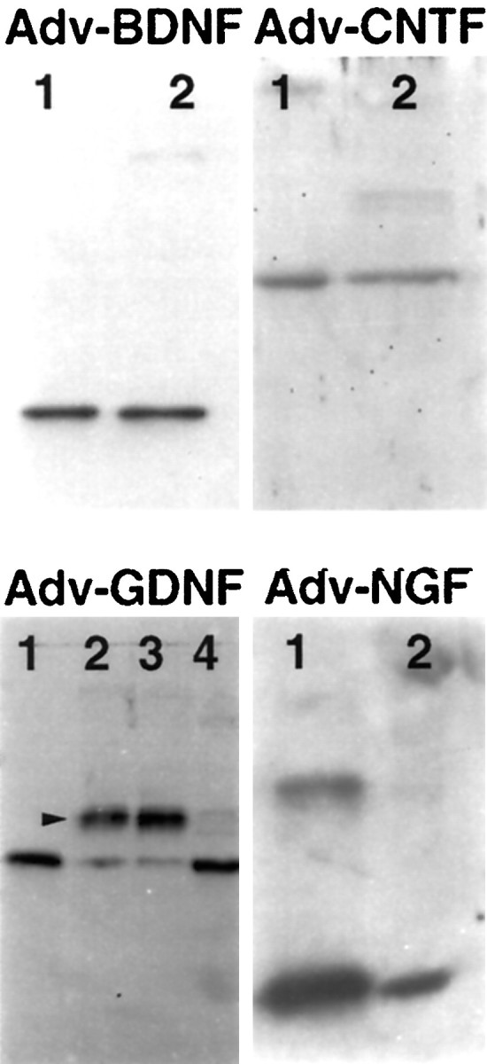
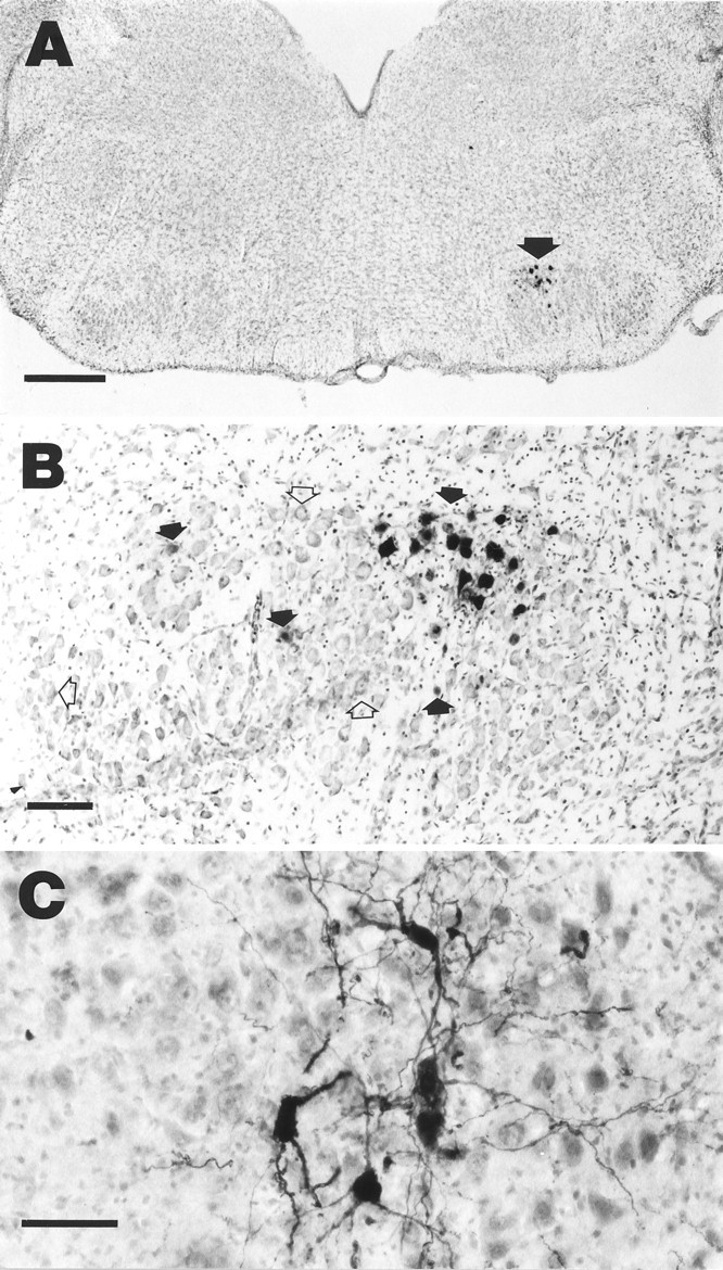
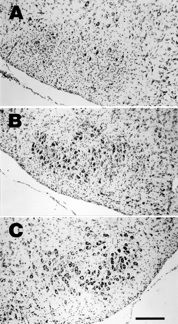
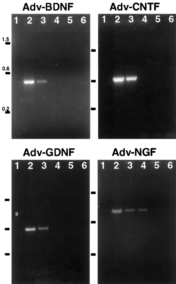
Application of neurotrophic factors (NFs) to the cut stump of motor nerves of neonatal rats confers neuroprotection from trauma-induced neuronal death. To test whether motoneurons are capable of responding to endogenously produced NFs, facial motoneurons were genetically modified in vivo to express several NFs and then tested for their response to peripheral nerve damage. Replication-defective adenoviral vectors [Adv. Rous sarcoma virus (RSV)-nf] representing three families of NFs were constructed that carried genes for brain-derived neurotrophic factor (BDNF), ciliary neurotrophic factor (CNTF), glial cell-derived neurotrophic factor (GDNF), and nerve growth factor. Media from cultured cells transduced with Adv. RSV-nf contained NFs that supported the survival of cultured chick sensory neurons in the same manner as recombinant NF standards. When Adv.RSV-nf or an adenoviral vector containing the beta-galactosidase gene (Adv.RSV-beta-gal) were injected into the facial muscles of neonatal rats the vectors were retrogradely transported to the facial nucleus where the NFs or beta-gal were expressed. A fraction (approximately 10%) of the neurons were transduced as demonstrated by reverse transcriptase-PCR, histochemistry, and immunocytochemistry. In the case of Adv.RSV-BDNF, Adv.RSV-CNTF, and Adv.RSV-GDNF, a significant portion of the facial nucleus neurons was protected, 16.5, 18.2, and 53.3%, respectively, from death after axotomy, showing that neurons are capable of transporting the Adv. RSV-nf, expressing the recombinant NF genes, and responding to the NFs. In the case of Adv.RSV-GDNF, a greater number of facial nucleus motoneurons survived than were transduced, indicating that neighboring untransduced neurons were protected by the GDNF expressed by the transduced neurons by a paracrine mechanism.


Figures

Fig. 1.
Fig. 1.
Expression of NFs by HeLa cells transduced with Adv.RSV-nf. Western blot analyses were performed on conditioned media or cell extracts from HeLa cells transduced with Adv.RSV-nf. The media and cell extract contained proteins that co-migrated (lanes 2) with the corresponding purified recombinant growth factors used as standards (lanes 1). The protein produced from cells transduced with Adv.RSV-GDNF showed cross-reacting bands (arrowhead) that migrated at higher apparent molecular weights than the unglycosylated standard. Glycosidase digestion of the conditioned medium from cells transduced with Adv.RSV-GDNF converted most of the high molecular weight protein to the size of the GDNF standard (lane 4), indicating that the protein was glycosylated by the HeLa cells. (Lane 3 is Adv.RSV-GDNF conditioned medium incubated in glycosidase reaction buffer without the addition of glycosidase.) Neither untransduced HeLa cells nor cells transduced with Adv.RSV-β-gal produced proteins that cross-reacted with the anti-NF antibodies (not shown). Molecular weights for the monomeric forms of recombinant standards are: rat CNTF, 23 kDa; human BDNF, 14 kDa; human β-NGF, 14 kDa; and human GDNF, 15 kDa.
Fig. 2.
Fig. 2.
Biological activity of NFs produced by HeLa cells transduced with Adv.RSV-nf vectors in vitro. Primary cultures of dissociated DRG (A) and NG (B) cells from chick embryos were grown in media containing culture medium from untransduced HeLa cells (white bar), 25% (v/v) of conditioned medium from HeLa cells (CM) transduced with Adv.RSV-β-gal (cross-hatched gray bars), or Adv.RSV-nf (black bars). In the case of CNTF a crude extract of cells transduced with Adv.RSV-CNTF or Adv.RSV-β-gal was mixed with standard medium. Surviving neurons were counted after 48 hr. *Value significantly different (p < 0.05) from Adv.RSV-β-gal value by one-way ANOVA with Bonferroni’s multiple-comparison test. Error bars indicate SD. B+G, BDNF and GDNF;B+C+N, BDNF, CNTF, and NGF.
Fig. 3.
Fig. 3.
Retrograde transport and expression of Adv.RSV-β-gal and Adv.RSV-CNTF. A, β-Galactosidase-positive neurons in the facial nucleus (arrow) 7 d after injection of Adv.RSV-β-gal into facial muscles. Scale bar, 500 μm. B, Detail of facial nucleus of a different rat than in A showing β-galactosidase-positive neurons (filled arrows) and nontransduced neurons (open arrows). Scale bar, 100 μm. C, Facial nucleus neurons that are immunopositive for CNTF in a field of nontransduced neurons stained with cresyl violet. Scale bar, 50 μm.
Fig. 4.
Fig. 4.
Protective effect of Adv.RSV-nf on axotomized facial nucleus neurons. Photomicrographs of cross-sections through the facial nucleus 9 d after injection of the ipsilateral facial muscles with Adv.RSV-β-gal (A) and Adv.RSV-GDNF (B) and 7 d after facial nerve transection. C, A typical nucleus contralateral to the side injected. Scale bar, 200 μm.
Fig. 5.
Fig. 5.
Detection of NF mRNA in regions of the facial nucleus after retrograde transport of Adv.RSV-nf.Adv.RSV-nf was injected into the facial muscles of neonatal rats, and total RNA was extracted from brainstem tissue after 7 d and analyzed by RT-PCR. Lane 1, No RNA;lane 2, positive control plasmid; lanes 3, 4, ipsilateral; lanes 5, 6, contralateral;lanes 3, 5; plus RT; lanes 4, 6, minus RT. Numbers to the left indicate sizes of markers in kilobase pairs.

References

    1. Akli S, Caillaud C, Vigne E, Stratford-Perricaudet LD, Poenaru L, Perricaudet M, Kahn A, Peschanski MR. Transfer of a foreign gene into the brain using adenovirus vectors. Nat Genet. 1993;3:224–228. - PubMed
    1. Baumgartner BJ, Barnes EM., Jr . Analysis of neuronal mRNA levels by quantitative reverse transcriptase-polymerase chain reaction. In: Wouterlood FG, editor. Neuroscience protocols, Module 4, Sec 20. Elsevier Science; Amsterdam: 1994. pp. 1–13.
    1. Chomczynski P, Sacchi N. Single-step method of RNA isolation by acid guanidinium thiocyanate-phenol-chloroform extraction. Anal Biochem. 1987;162:156–159. - PubMed
    1. Davidson BL, Allen ED, Kozarsky KF, Wilson JM, Roessler BJ. A model system for in vivo gene transfer into the central nervous system using an adenoviral vector. Nat Genet. 1993;3:219–223. - PubMed
    1. Davies AM. Neurotrophic factor bioassay using dissociated neurons. In: Rush RA, editor. Nerve growth factors. Wiley; New York: 1989. pp. 95–109.

MeSH terms

LinkOut - more resources