. 1997 Sep 1;17(17):6565-74.
doi: 10.1523/JNEUROSCI.17-17-06565.1997.

Kappa-opioid receptor activation modulates Ca2+ currents and secretion in isolated neuroendocrine nerve terminals

Affiliations

Kappa-opioid receptor activation modulates Ca2+ currents and secretion in isolated neuroendocrine nerve terminals

K I Rusin et al. J Neurosci. .

Abstract

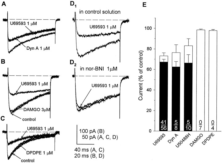
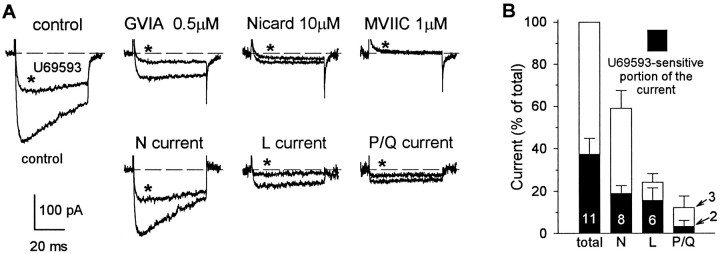
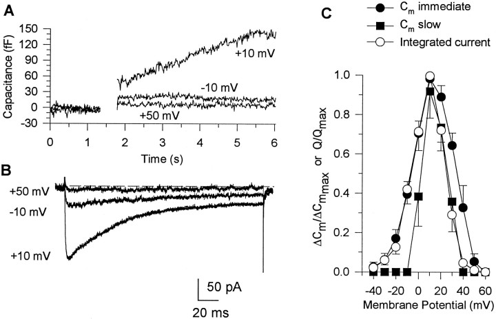
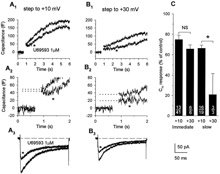
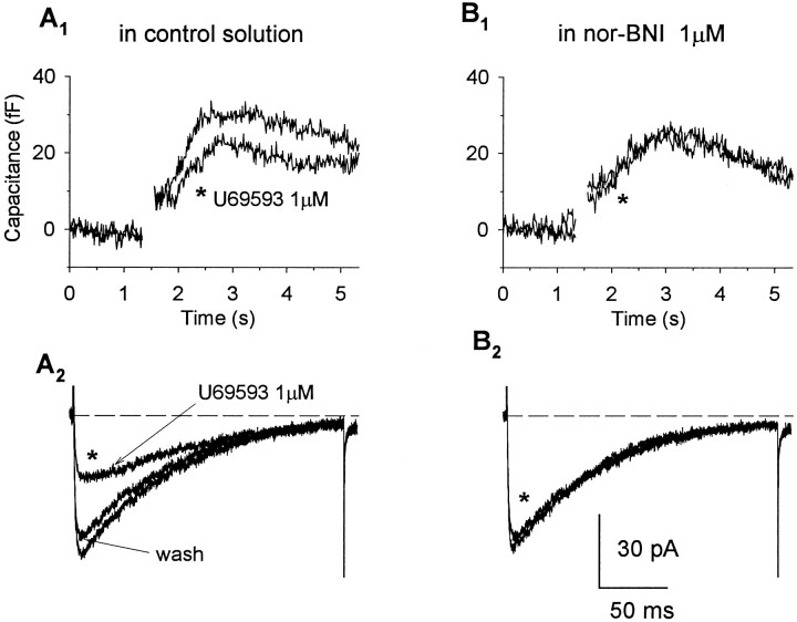
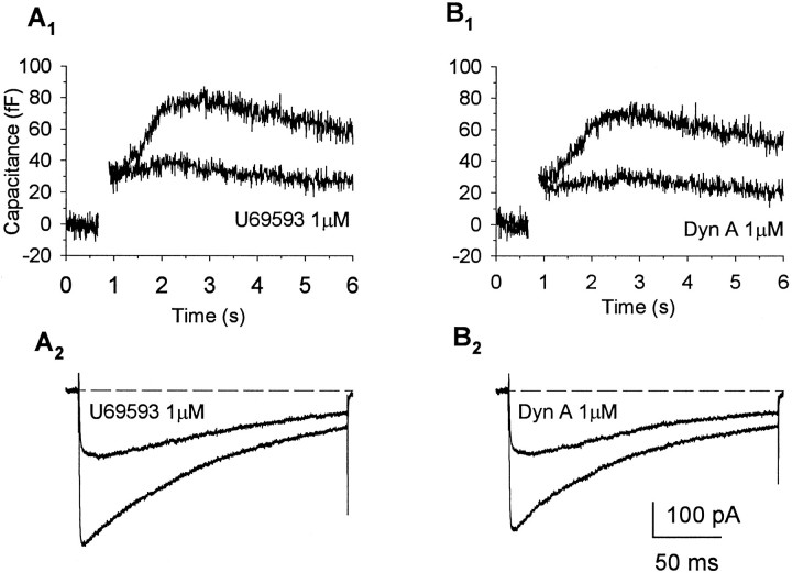
Whole-cell patch-clamp recordings were performed together with time-resolved measurements of membrane capacitance (Cm) in nerve terminals acutely dissociated from neurohypophysis of adult rats to investigate modulation of Ca2+ currents and secretion by activation of opioid receptors. Bath superfusion of the kappa-opioid agonists U69,593 (0.3-1 microM), dynorphin A (1 microM), or U50,488H (1-3 microM) reversibly suppressed the peak amplitude of Ca2+ currents 32. 7 +/- 2.7% (in 41 of 56 terminals), 37.4 +/- 5.3% (in 5 of 8 terminals), and 33.5 +/- 8.1% (in 5 of 10 terminals), respectively. In contrast, tests in 11 terminals revealed no effect of the mu-opioid agonist [D-Pen2,5]-enkephalin (1-3 microM; n = 7) or of the delta-agonist Tyr-D-Ala-Gly-N-Me-Phe-Gly-ol (1 microM; n = 4) on Ca2+ currents. Three components of high-threshold current were distinguished on the basis of their sensitivity to blockade by omega-conotoxin GVIA, nicardipine, and omega-conotoxin MVIIC: N-, L-, and P/Q-type current, respectively. Administration of U69,593 inhibited N-type current in these nerve terminals on average 32%, whereas L-type current was reduced 64%, and P/Q-type current was inhibited 28%. Monitoring of changes in Cm in response to brief depolarizing steps revealed that the kappa-opioid-induced reductions in N-, L-, or P/Q-type currents were accompanied by attenuations in two kinetically distinct components of Ca2+-dependent exocytotic release. These data provide strong evidence of a functional linkage between blockade of Ca2+ influx through voltage-dependent Ca2+ channels and inhibitory modulation of release by presynaptic opioid receptors in mammalian central nerve endings.


Figures

Fig. 1.
Fig. 1.
Opioid-induced inhibition of Ca2+ currents mediated via κ-opioid receptors.A–C, Recordings from three different terminals of whole-terminal Ca2+ currents evoked by a 100 msec (A, C) or 50 msec (B) step to +10 mV from −90 mV before the application and in the presence of selective κ-opioid (dynorphin A, 1 μm; U69,593, 1 μm), δ-opioid (DPDPE, 1 μm), or μ-opioid (DAMGO, 3 μm) agonists. Only the κ-opioid agonists reversibly inhibited Ca2+ currents. D1,D2, Recordings from another terminal showing that this effect of U69,593 was blocked by the κ-selective antagonist nor-BNI (1 μm). E, Comparison of effects of κ-, μ-, and δ-opioid-selective drugs on Ca2+ current. Open bars represent the mean ± SEM of the values obtained in the presence of a particular agonist (number of terminals shown in denominator); values are expressed as a percentage of the predrug control current. Current amplitudes were reduced significantly (p < 0.05) by U69,593, dynorphin A, and U50,488H but not by DAMGO and DPDPE. Filled barsrepresent only the data obtained from opioid-responsive terminals (number shown in numerator) and demonstrate equivalent inhibitory efficacy of the three κ-opioid agonists examined.
Fig. 2.
Fig. 2.
κ-Opioids inhibited GVIA-sensitive, nicardipine-sensitive, and MVIIC-sensitive Ca2+current components. A, upper, Recordings from a single nerve terminal show the effects of U69,593 (traces marked with asterisks) on Ca2+ currents evoked by 50 msec steps to +10 mV from a holding potential of −90 mV before the application and in the presence of Ca2+ channel-type selective blockers. Sequential administration of GVIA (0.5 μm), nicardipine (10 μm), and MVIIC (1 μm) completely abolished whole-terminal current. The ability of U69,593 to suppress Ca2+ current was greatly reduced after application of each of these blockers. A, lower, Subtraction of the corresponding currents evoked before and after application of antagonists yielded pharmacologically distinguished N-, L-, and P/Q-type current components in control conditions and in the presence of U69,593. B, Relative contribution of pharmacologically distinguished N-, L-, and P/Q-type current components to the total whole-terminal current and their sensitivity to the inhibitory effect of U69,593. Bars in the graph represent the mean ± SEM of the normalized current amplitudes obtained from the number of terminals shown, examined using the experimental protocol depicted in A.
Fig. 3.
Fig. 3.
Comparison of current–voltage and ΔCm–voltage relationships determined for single nerve endings. Cm responses (A) and corresponding Ca2+currents (B) evoked in a single terminal by 200 msec step depolarizations to different command potentials (shown near corresponding traces) from a holding potential of −90 mV. The depolarizations to −10 and +50 mV produced small currents that were accompanied by an immediate, step-likeCm increase, whereas steps to +10 mV evoked a larger current and prolonged Cm increase lasting for several seconds. Ca2+-dependentCm responses were corrected for transient ΔCm artifacts (as explained in text) by subtracting the response to a 5 msec depolarization aligned with the pulse. C. Integrated Ca2+ currents (open circles; n = 8) and corresponding immediate (filled circles;n = 8) and slow (filled squares; n = 6)Cm responses plotted against command potential at which they were evoked.
Fig. 4.
Fig. 4.
Activation of κ-opioid receptors attenuates immediate and slow Cm responses evoked by steps inducing smaller Ca2+ influx.Cm increases (A1,A2, B1,B2) and corresponding Ca2+currents (A3, B3) recorded in response to 200 msec depolarizations to +10 mV (A) and +30 mV (B) from a holding potential of −90 mV. Administration of U69,593 appreciably reduced Ca2+ influx (32.1%, from 18.53 to 12.58 pC) evoked by depolarization to +10 mV but had little effect on the corresponding immediate Cm response (9% reduction, from 49 to 44 fF, indicated by dotted lines). Depolarization to +30 mV induced much smaller Ca2+influx (9.1 pC) that was attenuated further by activation of κ-opioid receptors (to 6.23 pC, representing 31.5% blockade). Under this condition of reduced driving force for Ca2+ influx, inhibition of the immediate Cm response by the κ-opioid agonist was markedly increased (47%, from 36 to 19 fF). Opioid administration also reduced the slowly increasingCm component recorded in this nerve ending, and this effect of U69,593 was greater on the response measured at +30 mV (33.9%, from 118 to 78 fF) compared with that recorded at +10 mV (17.7%, from 192 to 158 fF). C, Bar graph summarizes the effects of U69,593 on the immediate step and slowly increasing components of Cm responses. Bars represent the mean ± SEM of the normalized Cmresponses pooled from κ-opioid-responsive terminals (number shown in numerator, out of the total number of terminals examined, shown in denominator) for immediate and slowCm responses evoked at command potentials of +10 or +30 mV, expressed as a percent of the corresponding predrug control values. Reduction of depolarization-evoked Ca2+ influx to submaximal levels resulted in an increased probability for modulation of the immediateCm response by κ-opioid receptor activation from 32% (13 of 41) at +10 mV to 100% (6 of 6) at +30 mV, without significantly (p = 0.19,NS indicates not significant) altering the magnitude of the inhibitory effect of U69,593 on these initial exocytotic events. In contrast, the inhibitory effect of U69,593 on the slowCm responses was significantly increased under conditions in which the driving force for Ca2+influx and the net amount of depolarization-evoked Ca2+ influx was reduced (asteriskindicates p < 0.001).
Fig. 5.
Fig. 5.
Activation of κ-opioid receptors attenuates depolarization-evoked exocytotic Cmresponses in neurohypophysial nerve endings in nor-BNI-sensitive manner. Cm increases (A1, B1) and corresponding Ca2+ currents (A2, B2) recorded in response to 200 msec depolarizations to +10 mV from a holding potential of −90 mV in control solution (A) and in the presence of the selective κ-opioid antagonist nor-BNI (B). Administration of U69,593 reduced the evoked Ca2+ current and accompanying slowly increasingCm response, and these effects were prevented after blockade of κ-opioid receptors by nor-BNI (1 μm).
Fig. 6.
Fig. 6.
Comparison of effects of U69,593 and dynorphin A demonstrates the similar efficacy of the two κ-opioid agonists in inhibiting Ca2+ currents and slowCm responses. Cmincreases (A1, B1) and corresponding Ca2+ currents (A2, B2) recorded in the same nerve terminal in response to 200 msec depolarizations to +10 mV from a holding potential of −90 mV in control solution and in the presence of U69,593 (A) and dynorphin A (B). Administration of U69,593 reduced the evoked Ca2+ current and accompanying slowly increasingCm response to a degree similar to that seen with dynorphin A.

Similar articles

Cited by

References

    1. Adamson P, Xiang JZ, Mantzourides T, Brammer MJ, Campbell IC. Presynaptic α2-adrenoceptor and κ-opiate receptor occupancy promotes closure of neuronal (N-type) calcium channels. Eur J Pharmacol. 1989;174:63–70. - PubMed
    1. Artalejo CR, Adams ME, Fox AP. Three types of Ca2+ channel trigger secretion with different efficacies in chromaffin cells. Nature. 1994;367:72–76. - PubMed
    1. Capogna M, Gahwiler BH, Thompson SM. Mechanism of μ-opioid receptor-mediated presynaptic inhibition in the rat hippocampus in vitro. J Physiol (Lond) 1993;470:539–558. - PMC - PubMed
    1. Cazalis M, Dayanithi G, Nordmann JJ. Hormone release from isolated nerve endings of the rat neurohypophysis. J Physiol (Lond) 1987;390:55–70. - PMC - PubMed
    1. Ciranna L, Feltz P, Schlichter R. Selective inhibition of high voltage-activated L-type and Q-type Ca2+ currents by serotonin in rat melanotrophs. J Physiol (Lond) 1996;490.3:595–609. - PMC - PubMed

Publication types