. 1997 Sep 1;17(17):6575-86.
doi: 10.1523/JNEUROSCI.17-17-06575.1997.

Adenoviral vector-mediated expression of B-50/GAP-43 induces alterations in the membrane organization of olfactory axon terminals in vivo

Affiliations

Adenoviral vector-mediated expression of B-50/GAP-43 induces alterations in the membrane organization of olfactory axon terminals in vivo

A J Holtmaat et al. J Neurosci. .

Abstract

B-50/GAP-43 is an intraneuronal membrane-associated growth cone protein with an important role in axonal growth and regeneration. By using adenoviral vector-directed expression of B-50/GAP-43 we studied the morphogenic action of B-50/GAP-43 in mature primary olfactory neurons that have established functional synaptic connections. B-50/GAP-43 induced gradual alterations in the morphology of olfactory synapses. In the first days after overexpression, small protrusions originating from the preterminal axon shaft and from the actual synaptic bouton were formed. With time the progressive formation of multiple ultraterminal branches resulted in axonal labyrinths composed of tightly packed sheaths of neuronal membrane. Thus, B-50/GAP-43 is a protein that can promote neuronal membrane expansion at synaptic boutons. This function of B-50/GAP-43 suggests that this protein may subserve an important role in ongoing structural synaptic plasticity in adult neurons and in neuronal membrane repair after injury to synaptic fields.


Figures
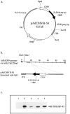
Fig. 1.
Fig. 1.
Construction of Ad-B-50/GAP-43 and Western blot analysis of vero cells expressing B-50/GAP-43 via Ad-B-50/GAP-43. a, Structure of pAdCMVB-50. The plasmid contains an expression cassette consisting of the human cytomegalovirus immediate early (CMV) promoter linked to the B-50/GAP-43 open reading frame (ORF) and the SV40 polyadenylation sequence [SV40 poly(A)] of the simian virus 40 early gene. The expression cassette was cloned between the inverted terminal repeat (ITR) (map units 0–1.25; the viral genome encapsidation signal) and the 9.2–15.5 map units (m.u.) sequence of the adenovirus type 5 genome. Furthermore, the plasmid contains the ampicillin resistance gene (AmpR) and an origin of replication (Ori). b, To generate the viral vector Ad-B-50/GAP-43, pAdCMVB-50 was linearized with SalI and transfected into producer 911 cells together with the ClaI and XbaI truncated Ad5dl309 genomic DNA. c, Western blot analysis detecting B-50/GAP-43 expression in Ad-LacZ-infected vero cells [5 × 10⁸ pfu (lane 1)], olfactory bulb (lane 2), and Ad-B-50/GAP-43-infected vero cells [5 × 10⁷ pfu (lane 3), 108 pfu (lane 4), 5 × 10⁸ pfu (lane 5)]. Vero cells were infected with the viral vectors for 1.5 hr, and after 48 hr the cells were harvested. Proteins extracted from these vero cells were separated by SDS-PAGE, blotted on nitrocellulose, and probed with anti-B-50/GAP-43 antibodies. A similarly treated sample of mouse olfactory bulb proteins was run (lane 2) as a reference sample. Note that Ad-LacZ-infected vero cells do not express B-50/GAP-43 and that B-50/GAP-43 from mouse olfactory bulb and B-50/GAP-43 expressed in vero cells via Ad-B-50/GAP-43migrate at the same position in the gel.
Fig. 2.
Fig. 2.
Ad-B-50/GAP-43- and Ad-LacZ-directed expression of B-50/GAP-43 and β-gal in mature olfactory neurons. A, B, Low power photomicrographs of transversal sections of mouse olfactory epithelia stained for β-gal (A) and B-50/GAP-43 (B) showing the patchy expression pattern of these molecules throughout the neuroepithelium 3 d after infusion of Ad-LacZ (A) and 12 d after infusion of Ad-B-50/GAP-43 (B). Note that areas containing high numbers of transduced cells (arrowheads in B) are alternated by areas without transduced cells (arrows in B).C–E, Confocal laser scanning micrographs of the expression of B-50/GAP-43 (C, D) and OMP (E) in olfactory neuroepithelia of Ad-LacZ (C) and Ad-B-50/GAP-43-injected mice (D, E) at 12 d after viral vector infusion. In mice infused with Ad-LacZ, B-50/GAP-43 expression is restricted to the normal population of immature cells in the basal portion of the epithelium (arrowhead in C). In contrast, in Ad-B-50/GAP-43-infused mice B-50/GAP-43-positive cells are detected in a scattered pattern through the olfactory neuroepithelium. Double-labeling for B-50/GAP-43 and OMP reveals numerous mature OMP-positive neurons coexpressing B-50/GAP-43 (arrows in D, E), indicating that mature neurons are efficiently transduced by Ad-B-50/GAP-43. Scale bar (shown in E): A, 500 μm;B, 125 μm; C–E, 50 μm.
Fig. 3.
Fig. 3.
B-50/GAP-43-induced morphogenic changes occur in mature OMP-positive olfactory neurons. Confocal laser scanning micrographs were taken from olfactory bulbs at 12 d after infusion of Ad-LacZ (A) or Ad-B-50/GAP-43 (B–D) and immunohistochemically stained for β-gal (A) or B-50/GAP-43 (B) or double-immunolabeled for OMP (C) and B-50/GAP-43 (D). Thin, long β-gal-expressing axons are present in a scattered pattern throughout the glomerulus and are often topped with small synaptic boutons (A, arrows). In contrast, B-50/GAP-43-expressing fibers exhibit large, morphologically altered axon endings that are formed predominantly at the edge of the glomerulus (B, arrows). Aberrant axon endings (arrowheads) are double-stained for B-50/GAP-43 (D) and OMP (C), indicating that these axon profiles represent morphologically changed mature olfactory nerve endings. Scale bar (shown in D): A, B, 25 μm;C, D, 30 μm.
Fig. 4.
Fig. 4.
B-50/GAP-43-overexpressing pri-mary olfactory axon endings grow toward the edge of glomeruli in the olfactory bulb. Confocal laser scanning micrographs were taken from mice infused with virus buffer (A) or with Ad-B-50/GAP-43 (B–E). Sections were stained with anti-B-50/GAP-43 antibodies at 3 (B), 5 (C), 8 (D), and 12 (A, E) d after infusion of Ad-B-50/GAP-43. B-50/GAP-43 is virtually absent in glomeruli in olfactory bulbs of control mice infused with virus buffer alone (A). At 3 and 5 d after infusion of Ad-B-50/GAP-43, B-50/GAP-43 is present in primary olfactory axons scattered throughout the neuropil of individual glomeruli (B, C), whereas at 8 and 12 d after infusion B-50/GAP-43-positive fiber endings become more and more located at the rim of individual glomeruli (D, E). This indicates that primary olfactory axon endings translocate from the glomerular neuropil to the edge of the glomerulus, where they form enlarged axonal terminals (arrows inD, E). Scale bar, 50 μm.
Fig. 5.
Fig. 5.
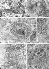
Immunoelectron microscopical analysis of mature primary olfactory axon profiles: coexpression of OMP and B-50/GAP-43 in axonal labyrinths. Ultrathin sections of Lowicryl-embedded olfactory bulbs of transgenic mice were labeled for B-50/GAP-43 (A–D) and OMP (E). A–C, Axon profiles (indicated by two arrowheads in C) that terminate in large concentrically organized membrane structures are always highly labeled for B-50/GAP-43 and are found predominantly in the vicinity of the glomerular border delineated by juxtaglomerular cells (indicated by jc in B andC). In adjacent sections the ultrastructurally abnormal axon endings were immunolabeled for B-50/GAP-43 (D) and OMP (E), indicating that axonal labyrinth formation occurs in mature OMP-positive olfactory axons overexpressing B-50/GAP-43. Scale bar (shown inE): A, 3.8 μm; B, 3.4 μm; C–D, 2.6 μm.
Fig. 6.
Fig. 6.
Various degrees of ultrastructurally altered axon profiles in transgenic mice. Ultrathin sections of Epon-embedded olfactory bulbs of a wild-type mouse (A) and transgenic littermate of L29 (B–E). In Epon-embedded olfactory bulbs, primary olfactory axon terminals have a relative electron-dense axoplasm (indicated by theasterisk in A), whereas dendritic profiles (indicated by d) exhibit an electron lucent appearance. Various degrees of ultrastructural alterations are present in glomeruli of transgenic mice, ranging from subtle (B) and moderate (C) to elaborate (D) axonal labyrinths. Some axon profiles exhibit a few sheaths of axolemma interspaced with axoplasm. The synaptic core elements contain small numbers of vesicles (arrow in C) compared with primary olfactory axon endings in wild-type mice (A). The elaborate axonal labyrinths consist of multiple layers of axolemma (E, higher magnification of square areain D) interspaced with ultrathin sheaths of axoplasm (a) and extracellular components (e). Note that the axonal labyrinth (D) occurs in close approximation with a glial cell process (indicated by gc) or in the vicinity of juxtaglomerular cell bodies (indicated by jc). Scale bar (shown in E): A–D, 1.5 μm;E, 0.02 μm.
Fig. 7.
Fig. 7.
Temporal dissection of morphological alterations in synaptic structures in Ad-B-50/GAP-43-infused mice. Ultrathin sections of Epon-embedded olfactory bulbs were taken from Ad-B-50/GAP-43- (A–E) or Ad-LacZ-injected (F) mice at 3 (A–C), 5 (D), and 12 (E, F) d after infusion of the viral vectors.A and B show examples of primary olfactory synapses that exhibit ultraterminal protrusions (arrows) arising from the synaptic bouton.C shows a synaptic bouton at 3 d with relatively advanced structural changes. Note that at 3 d the synaptic elements contain numerous vesicles and postsynaptic densities (indicated by pd in A). Furthermore, it can be clearly seen that the axolemmal extensions are continuous with the synaptic core (arrowhead in C). At 5 and 12 d, these structures have developed in axonal labyrinths with a synaptic core element virtually devoid of synaptic vesicles. Control axons expressing β-gal after transduction with Ad-LacZ are identified by preembedding labeling for β-gal. These axons exhibit a normal morphology. Scale bar (shown inF): A, D, E, F, 0.7 μm;B, 0.35 μm; C, 0.25 μm.

References

    1. Aigner L, Arber S, Kapfhammer JP, Laux T, Schneider C, Botteri F, Brenner H-R, Caroni P. Overexpression of the neural growth associated protein GAP-43 induces nerve sprouting in the adult nervous system of transgenic mice. Cell. 1995;83:269–278. - PubMed
    1. Akli S, Caillaud C, Vigne E, Stratford-Perricaudet LD, Poenaru L, Perricaudet M, Kahn A, Peschanski MR. Transfer of a foreign gene into the brain using adenovirus vectors. Nat Genet. 1993;3:224–228. - PubMed
    1. Aloyo VJ, Zwiers H, Gispen WH. Phosphorylation of B-50 protein by calcium-activated, phospholipid-dependent protein kinase and B-50 protein kinase. J Neurochem. 1983;41:649–653. - PubMed
    1. Bailey MS, Shipley MT. Astrocyte subtypes in the rat olfactory bulb; morphological heterogeneity and differential laminar distribution. J Comp Neurol. 1993;328:501–526. - PubMed
    1. Bajocchi G, Feldman SH, Crystal RG, Mastrangeli A. Direct in vivo gene transfer to ependymal cells in the central nervous system using recombinant adenovirus vectors. Nat Genet. 1993;3:229–334. - PubMed

Publication types

LinkOut - more resources