. 1997 Sep 1;17(17):6629-38.
doi: 10.1523/JNEUROSCI.17-17-06629.1997.

Developing neonatal rat sympathetic and sensory neurons differ in their regulation of 5-HT3 receptor expression

Affiliations

Developing neonatal rat sympathetic and sensory neurons differ in their regulation of 5-HT3 receptor expression

M Rosenberg et al. J Neurosci. .

Abstract

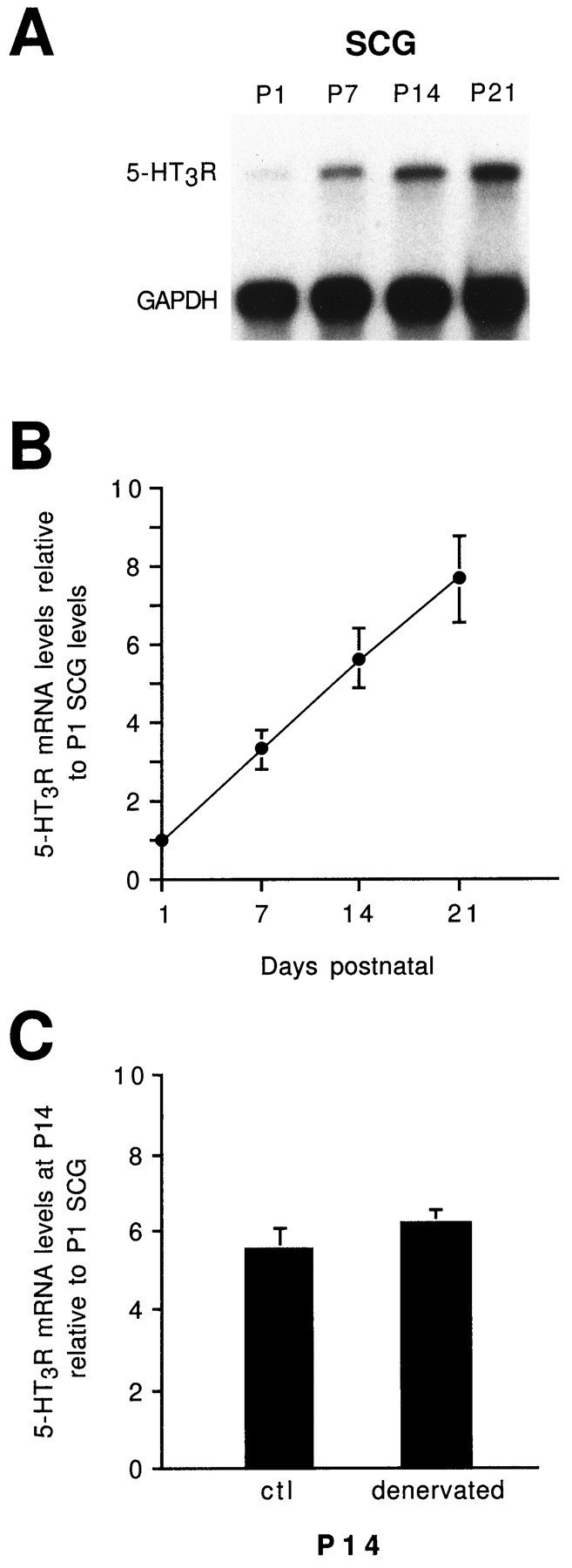
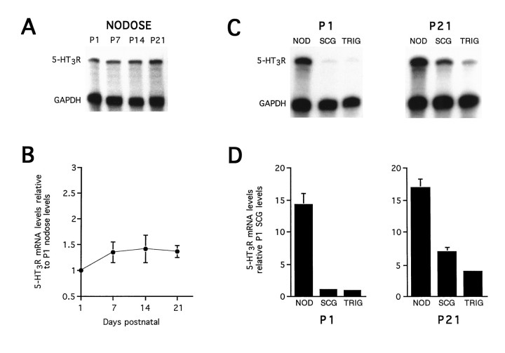
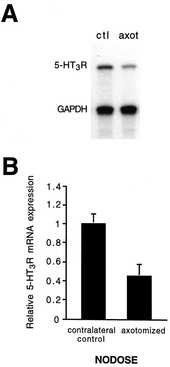
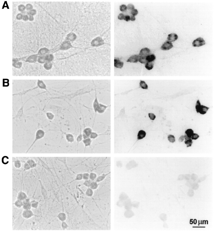
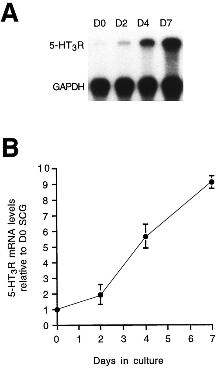
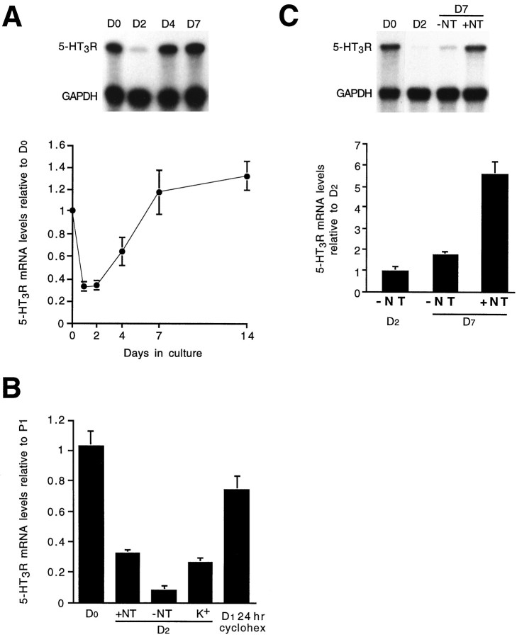
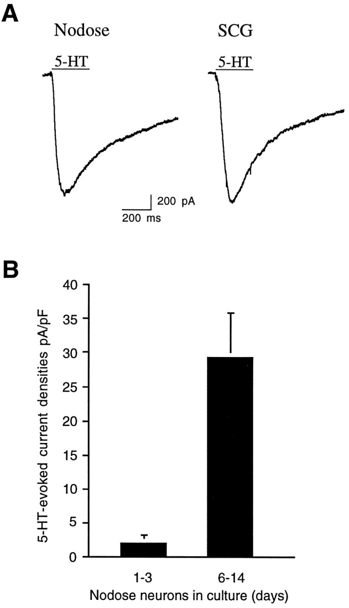
Serotonin 5-HT3 receptors (5-HT3Rs) are ligand-gated ion channels expressed by many peripheral neurons and are involved in several physiological processes. To learn more about the developmental regulation of 5-HT3R expression, we investigated rat sympathetic and vagal sensory neurons. We found that sympathetic and sensory neurons differ in their regulation of 5-HT3R expression during early postnatal life and as these neurons develop in culture. In SCG neurons 5-HT3R transcript levels are low at postnatal day 1 (P1) and increase 7.5-fold by P21; this increase occurs even after elimination of preganglionic innervation. In comparison, 5-HT3R mRNA levels in P1 nodose neurons are over 14-fold greater than in P1 SCG and change little by P21. We show that 5-HT3R transcript levels in nodose neurons depend on intact target innervation and drop by 60% after axotomy. When P1 SCG neurons develop in culture, we observed a significant increase in 5-HT3R expression: after 7 d in culture, transcript levels increase ninefold versus a threefold increase for neurons developing for 7 d in vivo. In contrast, 5-HT3R mRNA levels in cultured nodose neurons drop by 70% within 24 hr; however, this drop is transient. After 2 d, transcript levels begin to increase, and after 7 d, they are above initial values. We show that this delayed increase in 5-HT3R expression depends on neurotrophins. In both nodose and sympathetic neurons we found that the changes in 5-HT3R gene expression correlate directly with the appearance of 5-HT-evoked current densities.


Figures

Fig. 1.
Fig. 1.
Developmental increase in 5-HT3R mRNA expression in SCG. A, RNase protection assay for 5-HT3R and GAPDH mRNA expression in total RNA isolated fromP1, P7, P14, andP21 rat SCG. Total RNA (1 μg) was used for each reaction; the protected riboprobe sizes are 563 bases for 5-HT3R and 316 bases for GAPDH. GAPDH mRNA expression was assessed to ensure that equal amounts of total RNA were used in each reaction. B, Quantification of 5-HT3R mRNA expression in SCG: 5-HT3R expression increases 7.5-fold during the first 3 weeks of postnatal life. C, This figure shows that when P1 SCG ganglia are denervated for 2 weeks, there is no significant change in 5-HT3R mRNA expression, suggesting that innervation does not contribute to the observed developmental increase in 5-HT3R expression. The values represent the mean of six to eight experiments, and the error bars represent the SEM.
Fig. 2.
Fig. 2.
Differential 5-HT3R mRNA expression in SCG and nodose neurons. In situ hybridization with DIG-labeled antisense 5-HT3R probe on a section through P21 rat superior cervical ganglia (SCG), carotid artery (CA), nodose (NOD), and associated connective tissue. This figure is a montage of photomicrographs taken with a 20× objective and DIC optics on a Zeiss Axiovert 35 microscope. This figure shows that 5-HT3R mRNA is expressed at high levels in most nodose neurons and at lower levels in most SCG neurons and is not detectable in non-neuronal cells.
Fig. 3.
Fig. 3.
5-HT3R mRNA is highly expressed during early postnatal development in nodose ganglia. A, RNase protection assay for 5-HT3R mRNA expression performed as in Figure 1 on 1 μg of total RNA isolated from P1,P7, P14, and P21 rat nodose ganglia. B, Quantification of 5-HT3R mRNA expression in nodose: 5-HT3R expression changes little during the first 3 weeks of postnatal development. C, RNase protection assay depicting a comparison of 5-HT3R mRNA expression in 1 μg of total RNA prepared from P1and P21 nodose, SCG, and trigeminal ganglia.D, Quantification of 5-HT3R mRNA expression in nodose (NOD), SCG, and trigeminal ganglia (TRIG) at P1 andP21. The values represent the mean of six to eight experiments, and the error bars represent the SEM. In P1 SCG, P1 NOD, and P21 TRIG, the error bars are too small to resolve. This figure shows that atP1 5-HT3R expression is 14-fold higher in nodose than in SCG or trigeminal. However, in SCG and trigeminal, 5-HT3R mRNA expression increases significantly during the first 3 postnatal weeks such that by P21 the levels in nodose are only threefold larger than in SCG and fourfold larger than in trigeminal.
Fig. 4.
Fig. 4.
5-HT3R gene expression in nodose neurons depends on intact target innervation. A, RNase protection assay performed as in Figure 1 on 1 μg of total RNA prepared from P1 nodose ganglia that were axotomized 24 hr earlier (axot) and from control contralateral P1 nodose ganglia (ctl). B, Mean ± SEM 5-HT3R mRNA levels for axotomized P1 and P9 nodose ganglia are significantly different from control contralateral ganglia (p < 0.001; n = 12).
Fig. 5.
Fig. 5.
Most SCG and nodose neurons in culture express 5-HT3R transcripts. In situ hybridization with DIG-labeled riboprobe for 5-HT3R mRNA expression in P1 SCG and nodose neurons grown in culture for 7 d: A, SCG; B, nodose; C, control nodose neurons prehybridized with 400-fold excess unlabeled antisense 5-HT3R probe. Phase contrast (left) and DIC (right) photomicrographs were taken with a 40× objective on a Zeiss Axiovert 35 microscope. 5-HT3R mRNA is expressed by most D7 SCG and D7 nodose neurons.
Fig. 6.
Fig. 6.
Developmental expression of 5-HT3R transcripts by neonatal SCG neurons in culture. A, RNase protection assay performed as in Figure 1 on 1 μg of total RNA extracted from freshly dissociated SCG day 0 (D0) neurons and SCG neurons grown in culture for 2, 4, and 7 d.B, Quantification of 5-HT3R mRNA in cultured SCG neurons relative to D0. The values represent the mean of six to eight experiments, and the error bars represent the SEM.
Fig. 7.
Fig. 7.
5-HT3R mRNA levels decrease transiently in neonatal nodose neurons in culture. A, RNase protection assay and quantification performed as in Figure 1 on 1 μg of total RNA extracted from freshly dissociated nodose (day 0) neurons and nodose neurons grown in culture in the presence of NGF, BDNF, and NT-3 for 2, 4, 7, and 14 d. B, Quantification of 5-HT3R mRNA in cultured nodose neurons relative to P1: freshly dissociated (D0), with neurotrophins (+NT) for 48 hr, without neurotrophins (−NT) for 48 hr, with 40 mm K+ (K+) for 48 hr, or with cycloheximide for 24 hr (D1 24 hr cyclohex).C, Quantification of 5-HT3R mRNA in cultured nodose neurons relative to D2 measured at D7. Nodose neurons were grown for 2 d without neurotrophins (−NT) and then grown for 5 additional days with or without neurotrophins. 5-HT3R mRNA levels in nodose neurons after 5 d in neurotrophins were significantly greater (p< 0.001) than neurons grown in the absence of neurotrophins. The values represent the mean of three separate cultures, and each RNA sample was assayed twice; the error bars represent the SEM.
Fig. 8.
Fig. 8.
Nodose and SCG neurons express 5-HT-evoked currents. 5-HT-evoked currents were recorded from P1 nodose and SCG neurons grown in culture. A, Examples of 5-HT-evoked currents from D6 nodose and D7 SCG neurons. B, Mean 5-HT-evoked current densities (pA/pF) in nodose neurons grown for 1–3 d (n = 36) and 6–14 d (n = 25); the error bars represent the SEM.

References

    1. Andresen M, Kunze D. Nucleus tractus solitarius—gateway to neural circulatory control. Annu Rev Physiol. 1994;56:93–116. - PubMed
    1. Apud JA. The 5-HT3 receptor in mammalian brain: a new target for the development of psychotropic drugs? Neuropsychopharmacology. 1993;8:117–130. - PubMed
    1. Barthel L, Raymond P. Subcellular localization of α-tubulin and opsin mRNA in the goldfish retina using digoxigenin-labeled cRNA probes detected by alkaline phosphatase and HRP histochemistry. J Neurosci Methods. 1993;50:145–152. - PubMed
    1. Black IB. Regulation of autonomic development. Annu Rev Neurosci. 1978;1:183–214. - PubMed
    1. Chomczynski P, Sacchi N. Single-step method of RNA isolation by acid guanidium thiocyanate-phenol-chloroform extraction. Anal Biochem. 1987;162:156–159. - PubMed

Publication types

MeSH terms