. 1997 Sep 1;17(17):6685-96.
doi: 10.1523/JNEUROSCI.17-17-06685.1997.

Subunit composition, kinetic, and permeation properties of AMPA receptors in single neocortical nonpyramidal cells

Affiliations

Subunit composition, kinetic, and permeation properties of AMPA receptors in single neocortical nonpyramidal cells

M C Angulo et al. J Neurosci. .

Abstract

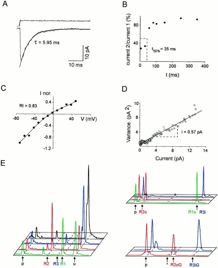
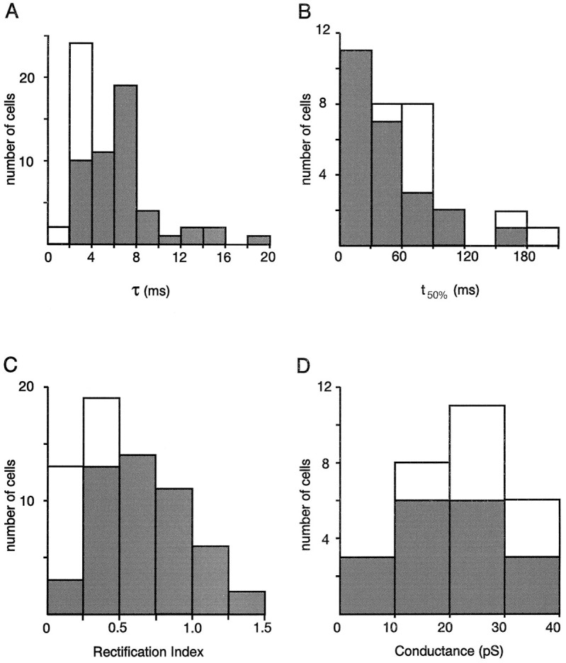
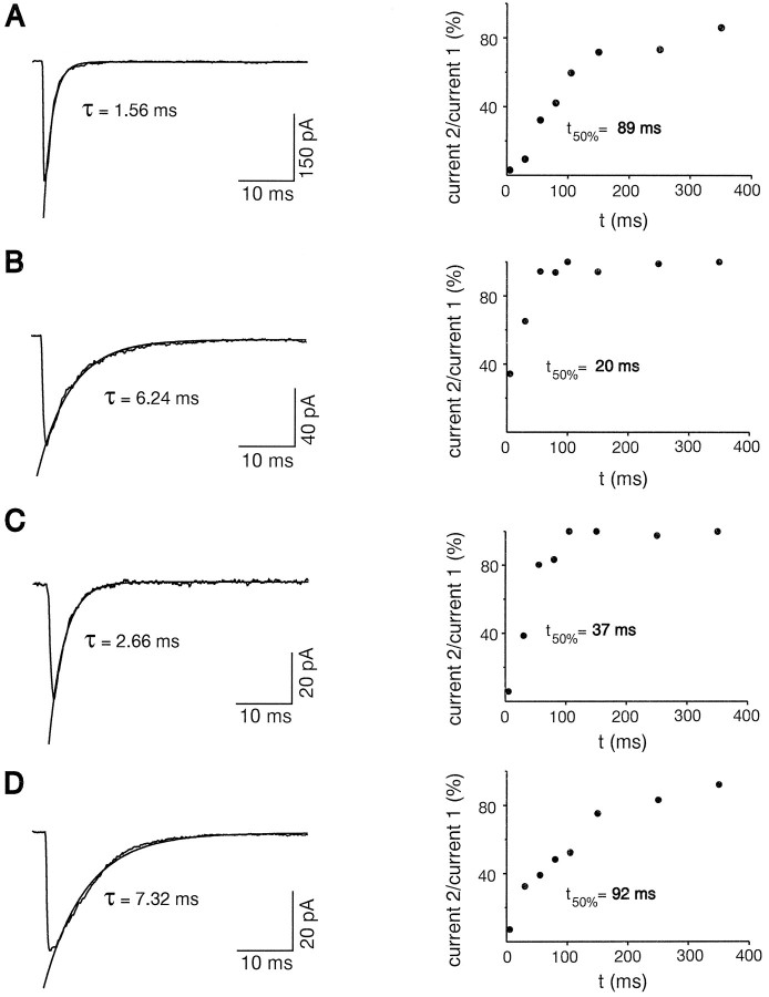
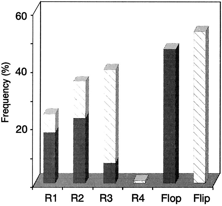
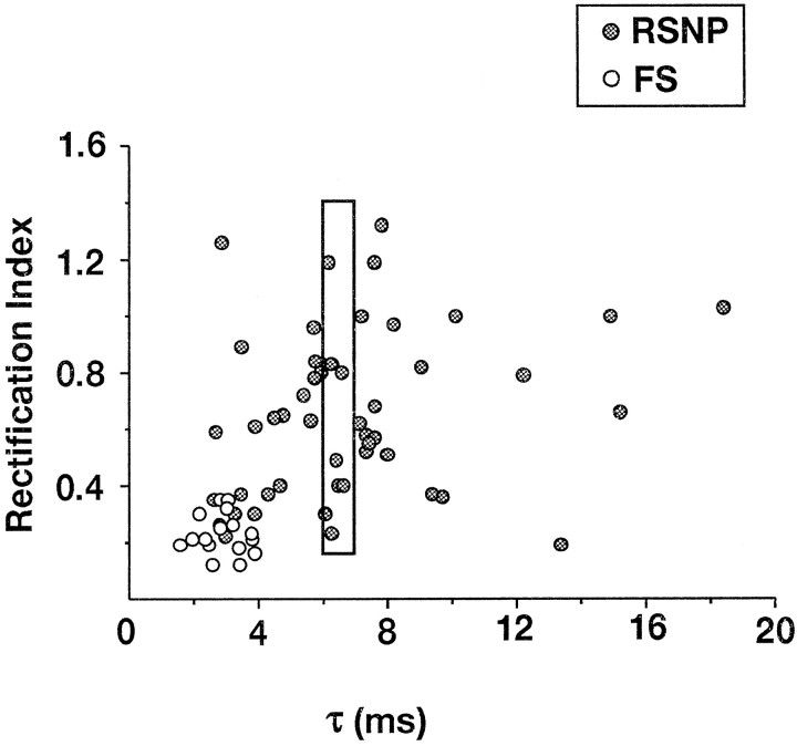
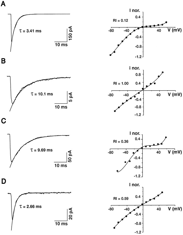
Native AMPA receptors (AMPARs) were investigated in neocortical fast-spiking (FS) and regular-spiking nonpyramidal (RSNP) cells. The onset of and recovery from desensitization as well as current rectification and single-channel conductance were studied by using fast glutamate application to outside-out patches. The GluR1-4 subunit, flip/flop splicing, and R/G editing expression patterns of functionally characterized cells were determined by single-cell reverse transcription-PCR to correlate the subunit composition of native AMPARs with their functional properties. Our sample, mostly constituted by RSNP neurons, predominantly expressed GluR3 flip and GluR2 flop. In individual cells, flip/flop splicing of each subunit appeared to be regulated independently, whereas for R/G editing all subunits were either almost fully edited or unedited. We confirmed that the relative GluR2 expression controls the permeation properties of native AMPARs, whereas none of the single molecular parameters considered appeared to be a key determinant of the kinetics. FS neurons displayed AMPARs with relatively homogeneous functional properties characterized by fast desensitization, slow recovery from desensitization, marked inward rectification, and large single-channel conductance. In contrast, these parameters varied over a wide range in RSNP neurons, and their combination resulted in various AMPAR functional patterns. Indeed, in different cells, fast or slow desensitization was found to be associated with either slow or fast recovery from desensitization. Similarly, fast or slow kinetics was associated with either strong or weak rectification. Our results suggest that kinetic and permeation properties of native AMPARs can be regulated independently in cortical neurons and probably do not have the same molecular determinants.


Figures

Fig. 1.
Fig. 1.
Functional and molecular analysis of AMPARs on a single RSNP neuron. Functional properties were investigated on an excised patch. Subsequently, a second whole-cell recording was obtained on the same cell, and the cellular content was harvested to investigate the GluR1–4 combination expressed. A, Averaged response (bottom trace) to 100 msec step applications (top trace) of 10 mm glutamate (holding potential, −72 mV). The current decay was fit by a single exponential function (τ = 5.95 msec) superimposed to the averaged current; average of 39 responses. B, Recovery from desensitization between currents induced by pairs of 1 msec pulses of glutamate separated by increasing time intervals (from 5 to 350 msec). The ratio of current 2/current 1 was plotted against time intervals. The time required for 50% recovery from desensitization was determined by interpolation (t50% = 35 msec). C,I–V curve of glutamate-induced responses. The averaged amplitudes of peak currents (averages of five responses) were measured at different potentials (from −72 to 48 mV) and normalized with respect to the peak current at −72 mV. Note the linear shape of the curve (RI = 0.83 andErev = −6 mV). The pointswere fit by a fourth-order polynomial function. D, Variance–amplitude plot obtained by nonstationary noise analysis of glutamate-induced responses (holding potential, −72 mV). Two-thirds of the plot was fit with a linear regression. The estimated single-channel current is obtained from the slope of the variance–amplitude relationship (i = 0.57 pA). The weighted mean single-channel conductance for AMPARs on this patch was 8.1 pS.E, Quantification of GluR1–4, flip/flop, and R/G edition proportions. GluR1–4 cDNA fragments were amplified with a fluorescent primer, cut with restriction enzymes, and submitted to capillary electrophoresis. The fluorescence profiles obtained are displayed with subunit-specific colors:green, GluR1; red, GluR2;blue, GluR3; black, GluR4. Time of migration is represented horizontally (fragment size increasing from left to right), and fluorescence intensity is represented vertically. Unincorporated primers peaks (p) are indicated.Left, GluR1–4 proportions. GluR1–4 cDNA fragments were coamplified and cut with subunit-specific enzymes. Positions of uncut (u; GluR1–4 cDNAs resistant to the specific enzyme) and cut cDNA fragments are indicated. No GluR4 was found in this cell. Thesmall peak present on all profiles at the same position (left of R2 peak) is attributable to a PCR artifact uncut by the enzymes. Top right panel, Flip/flop proportions of GluR1–4 subunits expressed. Subunit-specific amplifications were performed, followed by restriction with splice variant-specific enzymes. GluR1 and GluR3 were uncut (flop and flip, respectively), whereas GluR2 was cut (flop). Bottom right panel, R/G edition proportions. After subunit-specific amplification, products were cut with an editing variant-specific enzyme. Positions of edited flip (R3iG), edited flop (R2oG), and unedited (asterisk; not present but found in other analyzed cells) peaks are indicated. In this cell GluR3 flip and GluR2 flop were edited.
Fig. 2.
Fig. 2.
Functional properties of AMPARs in FS and RSNP neurons. Open and filled bars correspond to FS and RSNP cells, respectively. A, Desensitization time constants (τ; n = 66 cells).B, Recovery from desensitization (t50%; n = 32).C, Rectification index values calculated fromI–V curves (RI; n = 65). D, Weighted mean single-channel conductances (g; n = 28). Note that RSNP cells show wide distributions of the four functional parameters in comparison to FS cells.
Fig. 3.
Fig. 3.
Desensitization and recovery from desensitization of AMPARs in individual cells. Left, Averaged responses of excised patches elicited by step glutamate application. The current decay was fit by a single exponential function shown superimposed to the averaged current. Right, Plot of the ratio of current 2/current 1 against time, obtained by applying pairs of 1 msec pulses of glutamate separated by increasing time intervals (from 5 to 350 msec). A, Patch excised from an FS neuron. Note the fast desensitization (τ = 1.56 msec; average of six responses) and the slow recovery from desensitization (t50% = 89 msec). B, Patch excised from an RSNP neuron with a slow desensitization (τ = 6.24 msec; average of 11 responses) and a fast recovery from desensitization (t50% = 20 msec). C, Patch excised from an RSNP neuron with fast desensitization (τ = 2.66 msec; average of 10 responses) and fast recovery from desensitization (t50% = 37 msec). D, Patch excised from an RSNP neuron. Note the slow desensitization (τ = 7.32 msec; average of 25 responses) and the slow recovery from desensitization (t50% = 92 msec).
Fig. 4.
Fig. 4.
Relative abundance of GluR1–4 flip/flop in nonpyramidal neurons. Shown are mean proportions of GluR1–4 subunits found in 27 nonpyramidal neurons (23 RSNP cells and 4 FS cells). GluR2 and GluR3 mRNAs were expressed predominantly (GluR1, 24 ± 28%; GluR2, 36 ± 36%; GluR3, 39 ± 39%; GluR4, 1 ± 4%). Average percentages of flip (53 ± 43%) and flop (47 ± 43%) variants are indicated by hatched andfilled bars, respectively. The two barson the right represent the total percentages of flip and flop variants calculated from flip and flop proportions of each subunit.
Fig. 5.
Fig. 5.
Desensitization and rectification properties of AMPARs in FS and RSNP neurons. Desensitization time constants andRI were plotted for each cell. Open circles, FS neurons; filled circles, RSNP neurons. The points located inside therectangle show the large variability ofRI found for desensitization time constants of 6–7 msec.
Fig. 6.
Fig. 6.
Desensitization and rectification properties of AMPARs in individual cells. Left, Averaged responses of excised patches elicited by step glutamate application. The current decay was fit by a single exponential function shown superimposed to the averaged current. Right, I–V curves of glutamate-induced responses. The points were fit by third- or fourth-order polynomials. A, Patch excised from an FS neuron. Note the fast desensitization (τ = 3.41 msec; average of 35 responses) and the inward rectification of theI–V curve (RI = 0.12 andErev = −2 mV). B, Patch excised from an RSNP neuron. Note the slow desensitization (τ = 10.1 msec; average of 25 responses) and the linear I–V curve (RI = 1.00 and Erev = −6 mV). C, Patch excised from an RSNP neuron. Note the slow desensitization (τ = 9.69 msec; average of 11 responses) and the inward rectification of the I–V curve (RI = 0.36 and Erev = −5 mV). D, Patch excised from an RSNP neuron. Note the fast desensitization (τ = 2.66 msec; average of 10 responses) and the approximately linear I–V curve (RI= 0.59 and Erev = −5 mV).

References

    1. Bochet P, Audinat E, Lambolez B, Crépel F, Rossier J, Lino M, Tsuzuki K, Ozawa S. Subunit composition at the single-cell level explains functional properties of a glutamate-gated channel. Neuron. 1994;12:383–388. - PubMed
    1. Bowie D, Mayer ML. Inward rectification of both AMPA and kainate subtype glutamate receptors generated by polyamine-mediated ion channel block. Neuron. 1995;15:453–462. - PubMed
    1. Cauli B, Audinat E, Lambolez B, Angulo MC, Ropert N, Tsuzuki K, Hestrin S, Rossier J. Molecular and physiological diversity of cortical nonpyramidal cells. J Neurosci. 1997;17:3894–3906. - PMC - PubMed
    1. Colquhoun D, Jonas P, Sakmann B. Action of brief pulses of glutamate on AMPA/kainate receptors in patches from different neurones of rat hippocampal slices. J Physiol (Lond) 1992;458:261–287. - PMC - PubMed
    1. Connors BW, Gutnick MJ. Intrinsic firing patterns of diverse neocortical neurons. Trends Neurosci. 1990;13:99–104. - PubMed

Publication types

Substances

LinkOut - more resources