. 1997 Sep 1;17(17):6717-28.
doi: 10.1523/JNEUROSCI.17-17-06717.1997.

Differential expression of distinct members of Rho family GTP-binding proteins during neuronal development: identification of Rac1B, a new neural-specific member of the family

Affiliations

Differential expression of distinct members of Rho family GTP-binding proteins during neuronal development: identification of Rac1B, a new neural-specific member of the family

M L Malosio et al. J Neurosci. .

Abstract

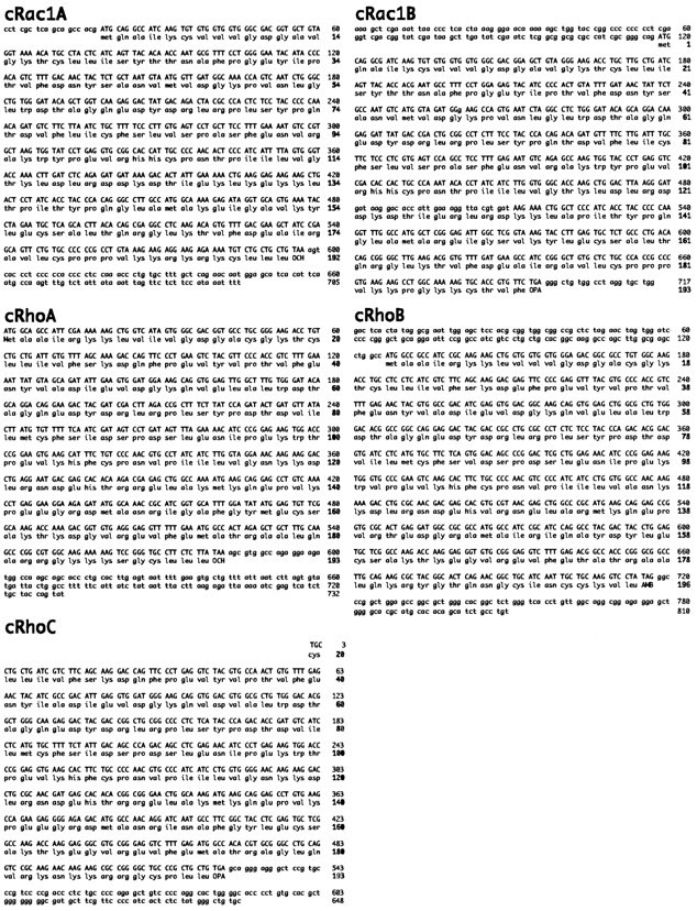
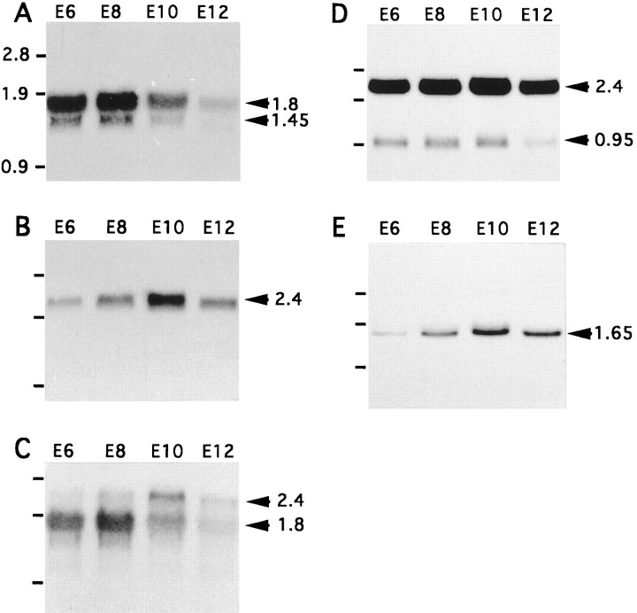
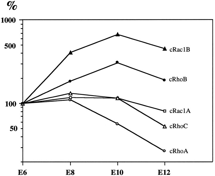
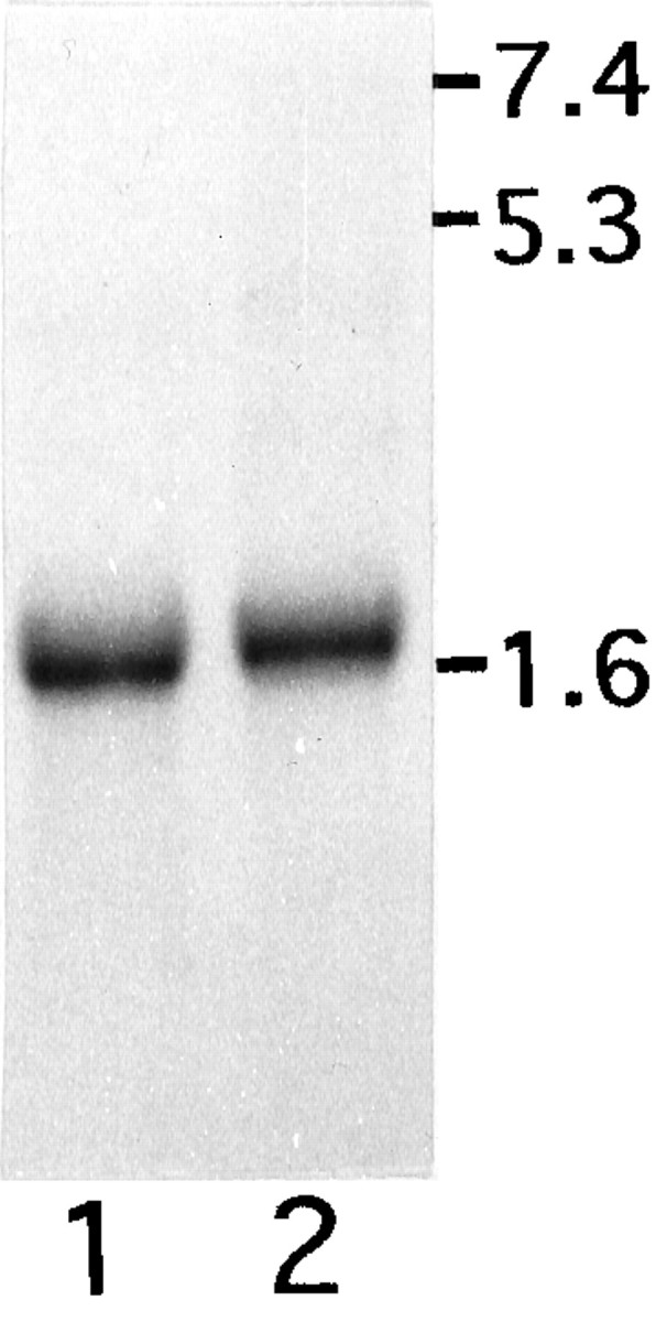
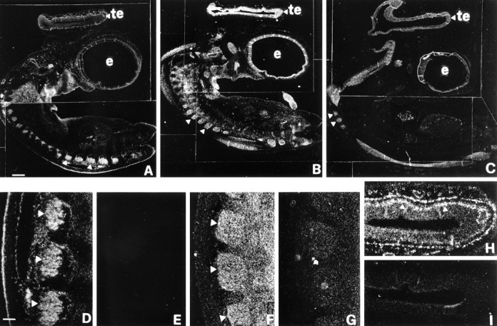
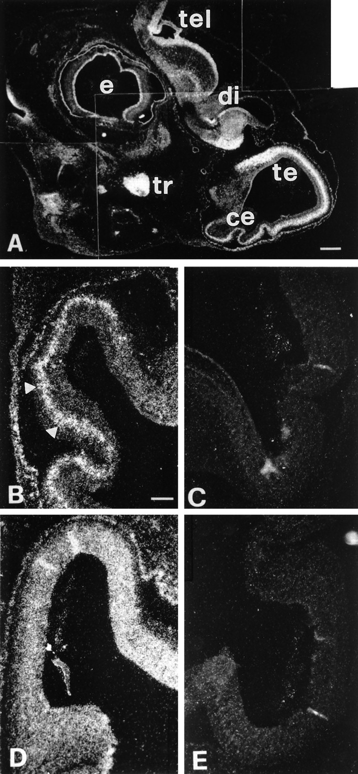
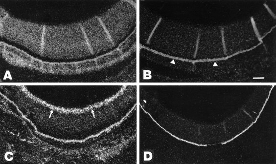
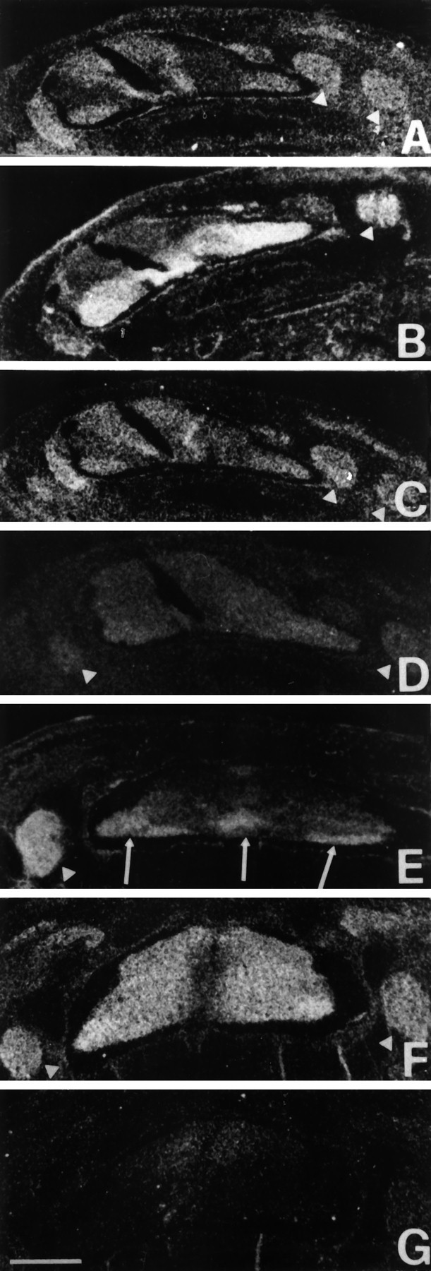
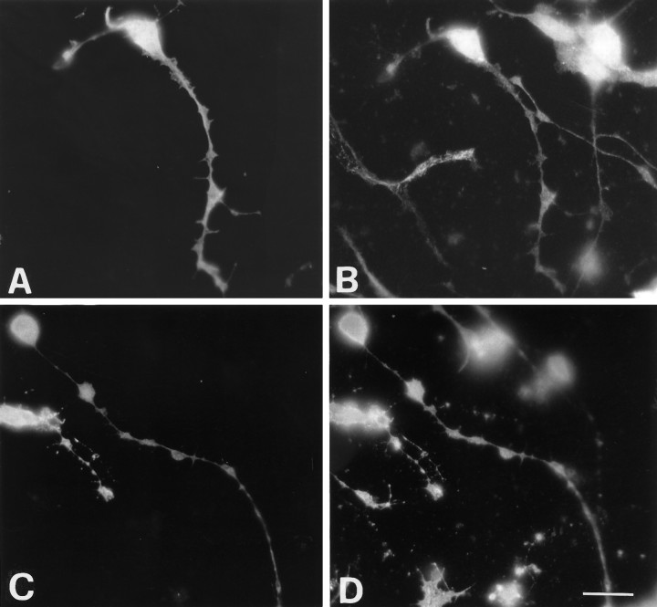
Previous studies on small GTP-binding proteins of the Rho family have revealed their involvement in the organization of cell actin cytoskeleton. The function of these GTPases during vertebrate development is not known. With the aim of understanding the possible role of these proteins during neuronal development, we have cloned and sequenced five members expressed in developing chick neural retinal cells. We have identified four chicken genes, cRhoA, cRhoB, cRhoC, and cRac1A, homologous to known human genes, and a novel Rac gene, cRac1B. Analysis of the distribution of four of the identified transcripts in chicken embryos shows for the first time high levels of expression of Rho family genes in the vertebrate developing nervous system, with distinct patterns of distribution for the different transcripts. In particular, cRhoA and cRac1A gene expression appeared ubiquitous in the whole embryo, and the cRhoB transcript was more prominent in populations of neurons actively extending neurites, whereas the newly identified cRac1B gene was homogeneously expressed only in the developing nervous system. Temporal analysis of the expression of the five genes suggests a correlation with the morphogenetic events occurring within the developing retina and the retinotectal pathway. Expression of an epitope-tagged cRac1B in retinal neurons showed a diffuse distribution of the protein in the cell body and along neurites. Taken as a whole, our results suggest important roles for ubiquitous and neural-specific members of the Rho family in the acquisition of the mature neuronal phenotype.


Figures

Fig. 1.
Fig. 1.
Nucleotide sequences of the chick Rho family GTPases cDNAs expressed in embryonic neural retina, and deduced primary sequences of the encoded polypeptides. The sequence data are available from GenBank under accession numbers U79757 (cRhoA),U79758 (cRhoB), U79759 (cRhoC), U79755(cRac1A), and U79756 (cRac1B).
Fig. 2.
Fig. 2.
Amino acid and nucleotide sequence comparisons.A, Amino acid sequence alignment of cRhoA, cRhoB, cRhoC, cRac1A, and cRac1B deduced polypeptides. Identical amino acids are shown in bold type. B, Percentages of identity between the coding regions of chick cDNAs cRhoA,cRhoB, cRhoC, cRac1A, andcRac1B and those of human cDNAs RhoA,RhoB, RhoC, Rac1, and Rac2.C, Percentages of amino acid identities between chick proteins cRhoA, cRhoB, cRhoC, cRac1A, and cRac1B and human proteins RhoA, RhoB, RhoC, Rac1, and Rac2.
Fig. 3.
Fig. 3.
Northern blot analysis of GTPase mRNA levels during neural retina development. Total RNA extracted from E6, E8, E10, and E12 neural retinas was electrophoresed on a 1% agarose gel and transferred to filters, as described in Materials and Methods. Filters were incubated with 32P-labeled probes specific forcRhoA (A), cRhoB(B), cRhoC(C), cRac1A(D), or cRac1B(E). RNA markers (in kilobases) are indicated to the left of each blot; the size of the transcripts (in kilobases) is indicated to the right.
Fig. 4.
Fig. 4.
Quantitation of the levels of expression of thecRhoA, cRhoB, cRhoC,cRac1A, and cRac1B transcripts during retina development. Quantitation was obtained by densitometry on autoradiograms from two experiments such as those shown in Figure 3. For cRhoA, cRhoC, andcRac1A transcripts, the values for both hybridizing bands were added (Fig. 3, A, C, and D, respectively). The values for the different GTPase transcripts were normalized to the values obtained from the corresponding 18 S ribosomal RNA hybridizations and plotted on a semilogarithmic scale. The E6 expression levels of the different transcripts were considered as 100%. Each value represents the mean obtained from two blots. Quantitation indicated that changes during neural retina development of the individual transcripts for cRhoA (Fig.3A) and cRac1A (Fig. 3D) were similar (quantitation not shown); on the other hand, the changes during development of the two transcripts of 2.4 and 1.8 kb recognized by the cRhoC probe (Fig. 3C) were different, showing highest levels of expression at E10 and E8, respectively (quantitation not shown).
Fig. 5.
Fig. 5.
Expression of cRac1B mRNA in neurons and glia from developing neural retina. Fifteen micrograms of total RNA extracted from cultures enriched in retinal glial cells (lane 1) or in retinal neurons (lane 2) prepared from E7 retinas were electrophoresed on a 1% agarose gel and transferred to filters, as described in Materials and Methods. Filters were incubated with a 32P-labeled probe specific forcRac1B. RNA markers (in kilobases) are indicated to theright.
Fig. 6.
Fig. 6.
In situ hybridization for different GTP-binding proteins of the Rho family in E6.5 chick embryos. Parasagittal sections were incubated with antisense probes forcRhoB (A), cRac1A(B), and cRac1B(C). Differences can be detected in the overall distribution of the mRNA for these three proteins. At this stage, the three different mRNAs were strongly expressed in the developing nervous system. DRGs show high levels of expression of the three mRNAs (arrowheads). The expression of cRhoB(D) and cRac1A(F) mRNAs in the DRGs is shown at higher magnification. E and G include similar fields from sections incubated with sense probes forcRhoB and cRac1A, respectively. In the tectum (te), cRac1A(B) and cRac1B(C) mRNAs show a homogeneous distribution. InH, a higher magnification of the area of the tectum shown in A reveals that cRhoB mRNA is strongly expressed in an external layer (arrowheads) corresponding to presumptive postmitotic neuroepithelial cells.I shows the area of the tectum from a control section incubated with a sense probe for cRhoB.e, Eye. Scale bars: A–C, 100 μm;D–I, 25 μm.
Fig. 7.
Fig. 7.
In situ hybridization for different GTP-binding proteins of the Rho family in E8.5 chick embryos.A, In situ hybridization ofcRhoB mRNA in parasagittal sections from E8.5 chick embryo head. Several structures of the developing nervous system show strong expression of cRhoB, including the retina in the eye (e), the telencephalon (tel), the diencephalon (di), the tectum (te), the cerebellum (ce), and the trigeminal ganglion (tr). B, Higher magnification of the developing cerebellum shows stronger expression ofcRhoB mRNA in the presumptive developing Purkinje cell layer (arrowheads). D, The developing cerebellum from a section similar to the one shown in Bshows a homogeneous expression of cRhoA mRNA. InC and E, sections incubated with sense probes for cRhoB and cRhoA, respectively, are shown as controls. Scale bars: A, 100 μm;B–E, 25 μm.
Fig. 8.
Fig. 8.
Expression of Rho GTPases in the developing chick retina. Expression of cRhoA (A, B) andcRhoB (C, D) mRNAs in the developing chick retina. Antisense (A, C) and sense (B, D) probes obtained from the respective cDNAs were incubated with sections from E8.5 chick embryos. Diffuse staining of the neural retina is observed for cRhoA (A), whereas stronger labeling is observed in the RGC layer (arrows) for cRhoB(C). The nonspecific signal given by the retinal pigmented epithelium is indicated by arrowheads inB. Scale bar, 25 μm.
Fig. 9.
Fig. 9.
Expression of Rho GTPases in the developing spinal cord. Sections including the spinal cord of E6.5 (A–D) and E8.5 (E–G) chick embryos were incubated with antisense probes forcRhoA (A), cRhoB(B, E), cRac1A (C), and cRac1B (D, F), and with a sense probe for cRac1B (G) as a control. Different patterns of expression can be observed for the different mRNAs. DRGs (arrowheads) can be observed on the sides of the spinal cord. In A–D the more oblique sections included two DRGs on one side of the spinal cord. InE, arrows indicate the localization ofcRhoB transcript in E8.5 spinal cord, mainly restricted to the motor neuron regions and to the floor plate (central arrow). Scale bar, 100 μm.
Fig. 10.
Fig. 10.
Distribution of cRac1B in cultured retinal neurons. Retinal neurons grown on laminin were transfected with the pcDNA-I-HA-Rac1B plasmid as described in Materials and Methods, and the cells were analyzed by immunofluorescence 24 hr after transfection. InA and B, cells were fixed with paraformaldehyde and permeabilized with 0.1% Triton X-100. InC and D, cells were fixed and permeabilized with cold (−20°C) methanol. Primary antibodies were monoclonal antibody against HA (A, C), polyclonal antibody against 200 kDa neurofilament protein (B), and polyclonal antibody against the integrin α6 subunit (D). Same fields are represented inA and B and in C andD). Scale bar, 10 μm.

References

    1. Adler R, Jerdan J, Hewitt AT. Responses of cultured neural retinal cells to substratum-bound laminin and other extracellular matrix molecules. Dev Biol. 1985;112:100–114. - PubMed
    1. Bartsch S, Husmann K, Schachner M, Bartsch U. The extracellular matrix molecule tenascin: expression in the developing retinotectal system and substrate properties for retinal ganglion cell neurites in vitro. Eur J Neurosci. 1995;7:907–916. - PubMed
    1. Biscardi JS, Cooper NGF, Maness PF. Phosphotyrosine-modified proteins are localized in Müller cells of the chick neural retina. Exp Eye Res. 1993;56:281–289. - PubMed
    1. Boussif O, Lezoualc’h F, Zanta MA, Mergny MD, Scherman D, Demeneix B, Behr JP. A versatile vector for gene and oligonucleotide transfer into cells in culture and in vivo: polyethylenimine. Proc Natl Acad Sci USA. 1995;92:7297–7301. - PMC - PubMed
    1. Cattelino A, Longhi R, de Curtis I. Differential distribution of two cytoplasmic variants of the α6β1 integrin laminin receptor in the ventral plasma membrane of embryonic fibroblasts. J Cell Sci. 1995;108:3067–3078. - PubMed

Publication types

MeSH terms

Grants and funding