

doi: 10.1073/pnas.94.17.8954.
Three functional luciferase domains in a single polypeptide chain
Affiliations
- PMID: 9256416
- PMCID: PMC22980
- DOI: 10.1073/pnas.94.17.8954
Item in Clipboard
Three functional luciferase domains in a single polypeptide chain
Proc Natl Acad Sci U S A.
.
Abstract
We report a unique case of a gene containing three homologous and contiguous repeat sequences, each of which, after excision, cloning, and expression in Escherichia coli, is shown to code for a peptide catalyzing the same reaction as the native protein, Gonyaulax polyedra luciferase (Mr = 137). This enzyme, which catalyzes the light-emitting oxidation of a linear tetrapyrrole (dinoflagellate luciferin), exhibits no sequence similarities to other luciferases in databases. Sequence analysis also reveals an unusual evolutionary feature of this gene: synonymous substitutions are strongly constrained in the central regions of each of the repeated coding sequences.
Figures
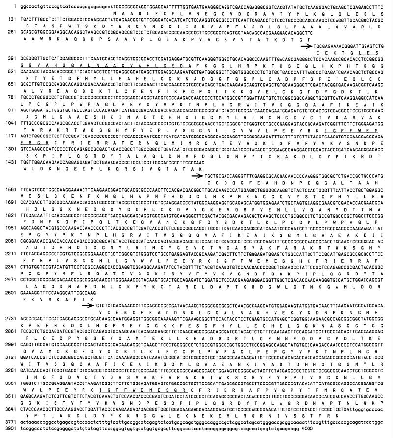
The complete nucleotide sequence of the pBS:LCF insert shown with its deduced translation aligned underneath. Lowercase letters represent 5′ and 3′ untranslated sequences. The arrows and offset sequences indicate the start of each of the three repeat regions. Sequences of trypsin peptides of purified G. polyedra LCF are underlined.
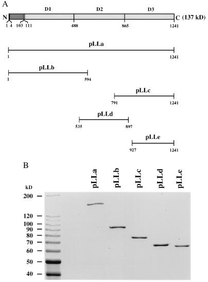
(A) Schematic diagram showing the expression constructs containing full-length (pLLa) or partial (pLLb, pLLc, pLLd, and pLLe) sequences of LCF. The amino acid numbers of each peptide are indicated. The expected molecular weights of GST–LCF fusion proteins are 164.6 kDa, 92.8 kDa, 75.8 kDa, 66.0 kDa, and 62.6 kDa for pLLa, pLLb, pLLc, pLLd, and pLLe, respectively. GST itself is 26 kDa. (B) Coomassie blue stained SDS/PAGE analyses of the GST–LCF fusion proteins. Expression and purification of GST–LCF proteins were performed as described. Protein standards (10-kDa ladder; BRL) were loaded onto the left lane; their molecular weights (in kDa) are indicated. About 1 μg of each protein was loaded per lane.
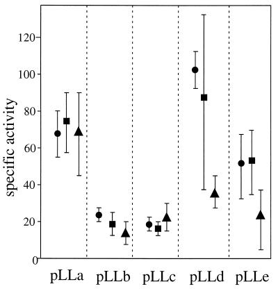
All five constructs were expressed and the proteins isolated in three separate runs; the average specific LCF activity values are shown for each of those runs. LCF activity assays were carried out as described; at least five measurements were made for each preparation, and the averages of initial maximum intensity were used for specific activity calculation (units, quanta·sec−1⋅μmol protein−1). Protein concentration of each GST–LCF was determined by Coomassie blue-stained SDS/PAGE; optical densities of bands were determined using nih image (version 1.60). Purified BSA (Calbiochem) was used as a standard and loaded onto the same gel in a series of known concentrations.
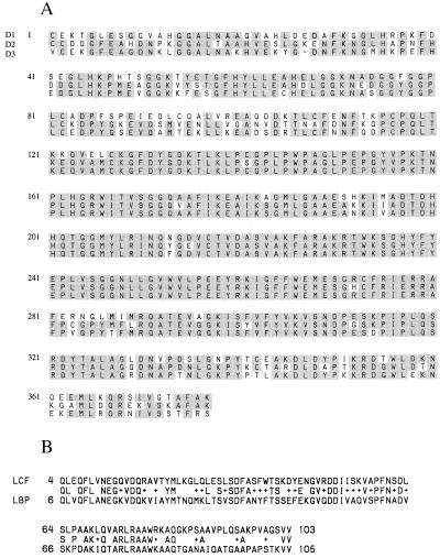
(A) Peptide sequence alignment of the three repeated domains, with the consensus residues shaded. The first residue of each repeated domain is numbered 1. (B) Peptide sequence alignment of the N-terminal regions of G. polyedra LCF and LBP. The identical residues are shown in the middle line (between LCF and LBP sequences). The symbol + indicates that the residues at that position are different but functionally conserved, based on the blast pam 240 program (23).
References
-
- Krasnow R, Dunlap J, Taylor W, Hastings J W, Vetterling W, Gooch V D. J Comp Physiol. 1980;138:19–26.
-
- Nicolas M-T, Morse D, Bassot J-M, Hastings J W. Protoplasma. 1991;160:159–166.
Publication types
MeSH terms
Substances
Associated data
- Actions
LinkOut - more resources
Full Text Sources
Other Literature Sources
Molecular Biology Databases
