. 1997 Aug 19;94(17):9063-8.
doi: 10.1073/pnas.94.17.9063.

Human argininosuccinate lyase: a structural basis for intragenic complementation

Affiliations

Human argininosuccinate lyase: a structural basis for intragenic complementation

M A Turner et al. Proc Natl Acad Sci U S A. .

Abstract

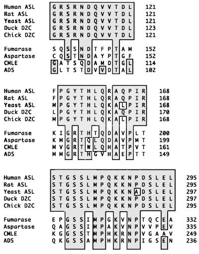
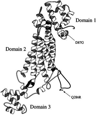
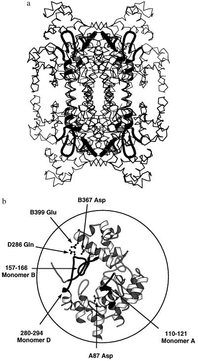
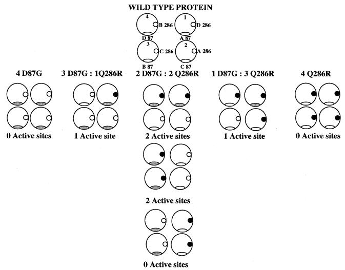
Intragenic complementation has been observed at the argininosuccinate lyase (ASL) locus. Intragenic complementation is a phenomenon that occurs when a multimeric protein is formed from subunits produced by different mutant alleles of a gene. The resulting hybrid protein exhibits enzymatic activity that is greater than that found in the oligomeric proteins produced by each mutant allele alone. The mutations involved in the most successful complementation event observed in ASL deficiency were found to be an aspartate to glycine mutation at codon 87 of one allele (D87G) coupled with a glutamine to arginine mutation at codon 286 of the other (Q286R). To understand the structural basis of the Q286R:D87G intragenic complementation event at the ASL locus, we have determined the x-ray crystal structure of recombinant human ASL at 4. 0 A resolution. The structure has been refined to an R factor of 18. 8%. Two monomers related by a noncrystallographic 2-fold axis comprise the asymmetric unit, and a crystallographic 2-fold axis of space group P3121 completes the tetramer. Each of the four active sites is composed of residues from three monomers. Structural mapping of the Q286R and D87G mutations indicate that both are near the active site and each is contributed by a different monomer. Thus when mutant monomers combine randomly such that one active site contains both mutations, it is required by molecular symmetry that another active site exists with no mutations. These "native" active sites give rise to the observed partial recovery of enzymatic activity.


Figures

Figure 1
Figure 1
The three regions of high sequence conservation among members of the ASL superfamily. ASL, argininosuccinate lyase; D2C, δ II crystallin; fumarase, E. coli fumarase C; aspartase, E. coli aspartase; CMLE, P. putida 3-carboxy-cis,cis-muconate lactonizing enzyme; ADS, B. subtilis adenylosuccinase.
Figure 2
Figure 2
A simulated annealing 2FoFo omit map for residues 158–161. All atoms displayed in the map were removed from the structure before a round of simulated annealing refinement. The map is contoured at 1 σ.
Figure 3
Figure 3
Schematic diagram showing the three-dimensional topology of an ASL monomer drawn with the program molscript (40). The amino acid side chains of residues D87 and Q287 have been drawn in a ball-and-stick representation. The three regions of highly conserved sequence are shaded in black.
Figure 4
Figure 4
Schematic diagrams showing the arrangement of the conserved regions (shaded in black) in (a) the tetramer and (b) the active site. The location of key residues are shown in a ball-and-stick representation. The residues are labeled according to the monomer on which they reside (A–D) and the residue number and type.
Figure 5
Figure 5
A pictorial representation of the active sites of the statistically available combinations of mutants in D87G:Q286R intragenic complementation of the ASL tetramer. For clarity the diagram has been drawn to show the interaction of only the D87 (ovals) and Q286 (circles) and the shading of the symbols represents the presence of the point mutations D87G and Q286R, respectively. Each large circle represents one of the four active sites found in the protein. In the schematic diagram of the native protein, residues 286 and 87 also are labeled according to which monomer they are found on. The monomers are designated A-D. Due to the molecular symmetry of the tetramer, in the case of the 2D87G:2Q286R tetramer there are three distinctly different ways of combining the monomers that will give rise to either two or zero native active sites being formed. For each combination of monomers, one also must consider which monomer contains the mutation, in the case of 3D87G:1Q286R tetramer, the Q286R mutation could be present on monomer A, B, C, or D, and this gives rise to four different possible solutions. The set of all possible combinations has a degeneracy corresponding to a binomial distribution of 1:4:6:4:1.

References

    1. Wistow G, Piatigorsky J. Science. 1987;236:1554–1556. - PubMed
    1. Piatigorsky J, O’Brien W E, Norman B L, Kalumuck K, Wistow G J, Borras T, Nickerson J M, Wawrousek E F. Proc Natl Acad Sci USA. 1988;85:3479–3483. - PMC - PubMed
    1. Yeh L-S, Elzanowski L T, Hunt L T, Barker W C. Comp Biochem Physiol. 1988;2:433–437. - PubMed
    1. Matsubasa T, Takiguchi M, Amaya Y, Matsuda I, Mori M. Proc Natl Acad Sci USA. 1989;86:592–596. - PMC - PubMed
    1. Woods S A, Miles J S, Guest J R. FEMS Microbiol Lett. 1988;51:181–186.

Publication types

Substances