. 1997 Aug 19;94(17):9119-24.
doi: 10.1073/pnas.94.17.9119.

mRNA binding protein mrnp 41 localizes to both nucleus and cytoplasm

Affiliations

mRNA binding protein mrnp 41 localizes to both nucleus and cytoplasm

D Kraemer et al. Proc Natl Acad Sci U S A. .

Abstract

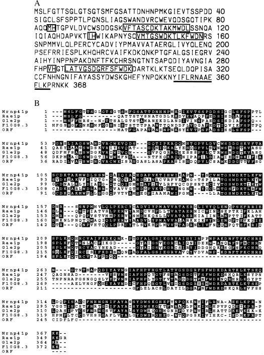
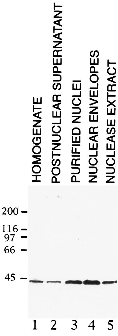
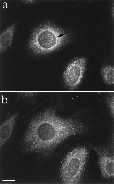
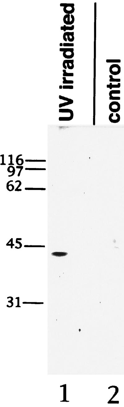
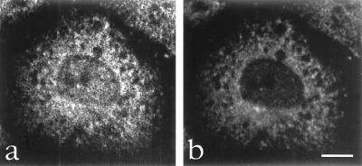
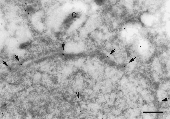
We have identified and molecularly characterized a human protein with a Mr of 40,880 Da. After UV irradiation of HeLa cells, this protein was cross-linked to poly(A)-containing mRNA and was therefore designated mrnp 41 (for mRNA binding protein of 41 kDa). Cell fractionation and immunoblotting showed mrnp 41 in both the cytoplasm and the nucleus and particularly in the nuclear envelope. Immunofluorescence microscopy localized mrnp 41 to distinct foci in the nucleoplasm, to the nuclear rim, and to meshwork-like structures throughout the cytoplasm. The cytoplasmic meshwork staining was disrupted by prior treatment of cells with the actin filament- or microtubule-disrupting drugs cytochalasin or nocodazole, respectively, suggesting association of mrnp 41 with the cytoskeleton. Double immunofluorescence with antibodies against mrnp 41 and the cytoplasmic poly(A) binding protein showed colocalization to the cytoplasmic meshwork. Immunogold electronmicroscopy confirmed mrnp 41's cytoplasmic and nucleoplasmic localization and revealed a striking labeling of nuclear pore complexes. Together these data suggest that mrnp 41 may function in nuclear export of mRNPs and/or in cytoplasmic transport on, or attachment to, the cytoskeleton. Consistent with a role of mrnp 41 in nuclear export are previous reports that mutations in homologs of mrnp 41 in Schizosaccharomyces pombe, designated Rae1p, or in Saccharomyces cerevisiae, designated Gle2p, result in mRNA accumulation in the nucleus although it is presently not known whether these homologs are mRNA binding proteins as well.


Figures

Figure 1
Figure 1
DNA-deduced amino acid sequence of human mrnp 41 and alignment with homologs. (A) Three repeats of Try-Asp (WD) motifs are boxed; the amino acid sequence retrieved from peptide sequencing is underlined. (B) Alignment of amino acid sequences of mnrp 41 and putative homologs in S. pombe (Rae1p), S. cerevisiae (Gle2p), C. elegans, and A. thaliana. Alignment was by the program lasergene navigator (DNAstar, Madison, WI).
Figure 2
Figure 2
Cell fractionation and Immunoblot with anti-mrnp 41 antibodies. A rat liver homogenate (lane 1) was fractionated by differential centrifugation into crude nuclei and a postnuclear supernatant (lane 2). Purified nuclei (lane 3) were fractionated by nuclease digestion yielding a nuclease extract (lane 5) and a nuclear envelope fraction (lane 4). Aliquots containing 25 μg of protein were subjected to SDS/PAGE. Proteins were transferred to nitrocellulose and immunoblotted using affinity-purified rabbit antibodies against mrnp 41.
Figure 3
Figure 3
Localization of mrnp 41 in HeLa cells by confocal immunofluorescence microscopy. HeLa cells were grown to subconfluency on a coverslip, permeabilized and fixed with methanol, and then probed with affinity-purified mouse anti-mrnp 41 antibodies (see Materials and Methods). An optical section corresponding to the equatorial plane of the nucleus shows staining of intranuclear foci, of the nuclear rim (see arrow head), and of a coarse meshwork (a). An optical section close to coverslip shows in addition a fine meshwork-like staining in the cytoplasmic periphery (b). (Bar = 10 μm.)
Figure 4
Figure 4
UV cross-linking of mrnp 41 to poly(A)-containing mRNA. Poly(A)-containing mRNA was isolated from HeLa cells that were either UV-irradiated (UV irradiated) or not UV-irradiated (control). The poly(A)-containing mRNA was digested with ribonucleases, the digest separated by SDS/PAGE, and the resolved proteins probed with affinity-purified mouse anti-mrnp 41 antibodies. Numbers to the left indicate Mr of marker proteins.
Figure 5
Figure 5
Double immunofluorescence confocal microscopy using mouse mAb against cPABP and affinity-purified rabbit antibodies against mrnp 41. Methanol-permeabilized and fixed HeLa cells were incubated with affinity-purified rabbit anti-mrnp 41 antibodies (a) and with monoclonal anti-cPAPB antibodies (b), and the bound antibodies were visualized with fluorescently labeled secondary antibodies. (Bar = 10 μm.)
Figure 6
Figure 6
Immunogold electron microscopy of HeLa cells using anti-mrnp 41 antibodies. Frozen ultrathin sections of HeLa cells were incubated with affinity-purified mouse anti-mrnp 41 antibodies and binding was detected with 10 nm of gold-coupled secondary antibodies. N, nucleoplasm; C, cytoplasm. Arrows point to gold particles at nuclear pore complexes: (Bar = 300 nm.)

References

    1. Daneholt B. Cell. 1997;88:585–588. - PubMed
    1. Piñol-Roma S, Dreyfuss G. Trends Cell Biol. 1993;3:151–155. - PubMed
    1. Piñol-Roma S, Dreyfuss G. Nature (London) 1992;355:730–735. - PubMed
    1. Visa N, Alzhanova-Ericsson A T, Sun X, Kiseleva E, Björkroth B, Wurtz T, Daneholt B. Cell. 1996;84:253–264. - PubMed
    1. Görlach M, Burd C C, Dreyfuss G. Exp Cell Res. 1994;211:400–407. - PubMed

Publication types

MeSH terms

Substances

Associated data