. 1997 Aug 19;94(17):9159-64.
doi: 10.1073/pnas.94.17.9159.

Expression of Sonic hedgehog gene in regenerating newt limb blastemas recapitulates that in developing limb buds

Affiliations

Expression of Sonic hedgehog gene in regenerating newt limb blastemas recapitulates that in developing limb buds

Y Imokawa et al. Proc Natl Acad Sci U S A. .

Abstract

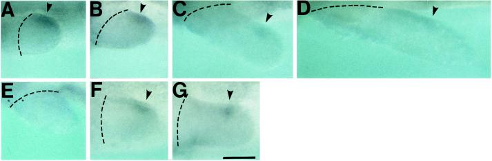
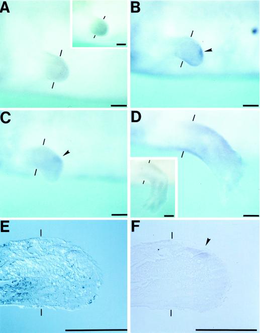
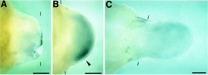
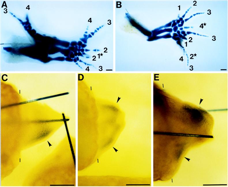
This study aimed at characterizing the Sonic hedgehog (shh) gene in newt limbs, which encodes a signaling molecule of the zone of polarizing activity (ZPA) responsible for determining the anterior-posterior axis of the embryonic chicken and mouse limbs. The reverse transcription-PCR showed that adult newt regenerating limbs express shh genes. In situ hybridization experiments demonstrated that shh genes were expressed in mesenchymal cells of the posterior region of both embryonic buds and regenerating blastemas of newt limbs, strongly suggesting the presence of ZPA in these tissues. Experiments of the axial reversal graft of blastemas further supported this suggestion. The grafted blastemas regenerated supernumerary limbs, and this has been explained by three models: the polar coordinate model, the boundary model, and the polarizing zone model. In favor of the third model, the shh gene was expressed not only in the original region (new anterior region) of the graft, but also ectopically in the other region (new posterior region) of the same graft. This study implies that the regenerating limb blastema produces ZPA as the signaling center of the AP patterning as in the developing limb bud and, therefore, supports the notion that the limb regeneration recapitulates the limb development.


Figures

Figure 1
Figure 1
Expression of shh in developing forelimb buds. Hybridization was performed with antisense probes (AD, F, and G) or with sense probes (E). (A) Stage 38, an early limb bud. (B, E, and F) Stage 40, a medium limb bud. (C and G) Stage 42, a late limb bud. (D) Stage 45 when two to three digits were formed. (AE) Views from the dorsal side of the embryos. (F and G) Views from the lateral side of the embryos. (Upper) Posterior region. shh-expressing region is shown by arrowheads. Broken lines indicate the base of limb buds. (Scale bar = 200 μm.)
Figure 2
Figure 2
Expression pattern of shh in larval regenerating limbs. Amputation planes are shown by thin bars. (AD) Blastemas of larval forelimbs viewed from the lateral side of the body. (Upper) Posterior region. (A) Four days after amputation, early to medium bud stage. (Inset) A case with sense probes. (B) Six days after amputaton, late bud stage. (C) Eight days, late bud to palette stage. (D) Twelve days after amputation, a 4-digit limb. (Inset) A case with sense probes. (E and F) A larval forelimb blastema at 6 days after amputation was treated for the in situ hybridization as above and was sectioned 10 μm thick. The sections were viewed through a microscope with (E) or without (F) Nomarski. (Upper) Posterior region. shh-expressing region is shown by arrowheads. (Scale bar = 200 μm.)
Figure 3
Figure 3
Expression pattern of shh in adult regenerating limbs. Amputation planes are shown by thin bars. Blastemas of adult forelimbs were viewed from the dorsal side. (Lower) Posterior region. (A) Fourteen days after amputation, dedifferentiation to early bud stage. (B) Twenty-one days after amputation, medium bud stage. (C) Twenty-eight days after amputation, palette stage. shh-expressing region is shown by an arrowhead. (Scale bar = 500 μm.)
Figure 4
Figure 4
Supernumerary limb formation and duplication of shh expression in regenerates of axial-reversed blastemas. (A and B) Skeletal patterns of supernumerary limbs. Arabic numerals show the number of digits. (Lower) Posterior region. (A) APDV axial reversal grafts. Light limbs of adult newts were amputated at a mid-upper arm level and the resulting blastemas were amputated at medium bud stage. Blastemas were rotated 180° to make the APDV axes the opposite of the axes of the original site and ipsilaterally grafted to the original stumps. 1, digit 1, which is partly hidden by the lower digit 2. Only part of its proximal phalanx can be seen. (B) AP axial reversal grafts. Right and left limbs were amputated at a mid-upper arm level and the resulting left blastemas at medium bud stage were contralaterally grafted to the light stumps without rotation. 2, another digit 2, which is not visible here because it is completely hidden by the lowest digit 3. The tip of 4 was found to be forked when it was seen through a stereoscopic microscope and, therefore, digit 4 is digit 4, which was formed by a fusion of two digits. (CE) Whole-mount in situ hybridization. (Lower) Posterior region. Amputation planes are shown by thin bars. (C) Control grafts. Control experiments for axial reversal grafting. Experiments were carried out as in A except that the blastemas were grafted without rotation to make the APDV axes of the graft meet those of the stump. Limbs were analyzed for shh expression 14 days after grafting. (D and E) Axial reversal grafts. (D) APDV axial reversal grafts. Limbs were subjected to the APDV axial reversal graft as described in A and were analyzed for shh expression at 14 days after grafting. (E) AP axial reversal grafts. Limbs were subjected to the AP axial reversal graft as described in B and were analyzed for shh expression 12 days after grafting. shh-expressing regions are shown by arrowheads. Solid bars in C and E are tungsten needles inserted to fix the grafts on stumps. (Scale bar = 500 μm.)

References

    1. Wallace H. Vertebrate Limb Regeneration. Toronto: Wiley; 1981.
    1. Stocum D L. Molecular Biology Intelligence Unit: Wound Repair, Regeneration and Artificial Tissues. New York: Springer; 1995.
    1. Tabin C J. Cell. 1991;66:199–217. - PubMed
    1. Riddle R D, Johnson R L, Laufer E, Tabin C. Cell. 1993;75:1401–1416. - PubMed
    1. Tickle C, Eichele G A. Rev Cell Biol. 1994;10:121–152. - PubMed

LinkOut - more resources