

Molecular evolution of integrins: genes encoding integrin beta subunits from a coral and a sponge
- PMID: 9256456
- PMCID: PMC23098
- DOI: 10.1073/pnas.94.17.9182
Molecular evolution of integrins: genes encoding integrin beta subunits from a coral and a sponge
Abstract
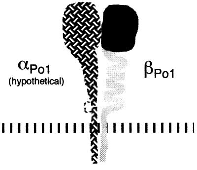
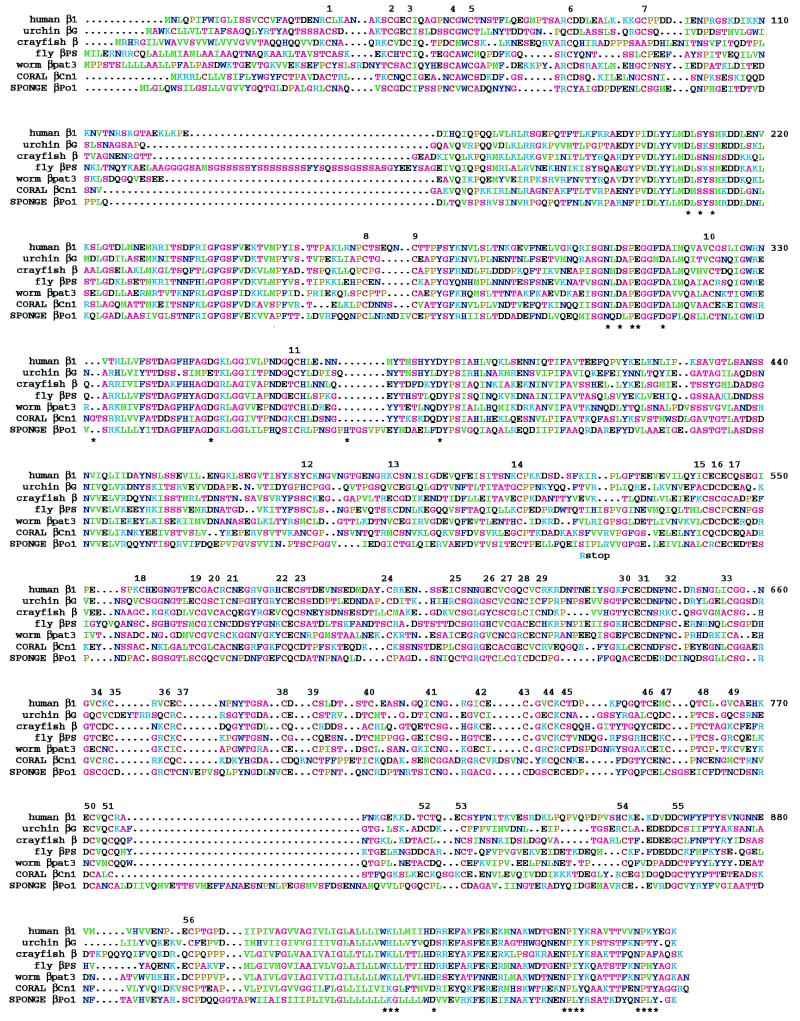
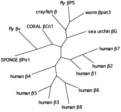
The integrin family of cell surface receptors is strongly conserved in higher animals, but the evolutionary history of integrins is obscure. We have identified and sequenced cDNAs encoding integrin beta subunits from a coral (phylum Cnidaria) and a sponge (Porifera), indicating that these proteins existed in the earliest stages of metazoan evolution. The coral betaCn1 and, especially, the sponge betaPo1 sequences are the most divergent of the "beta1-class" integrins and share a number of features not found in any other vertebrate or invertebrate integrins. Perhaps the greatest difference from other beta subunits is found in the third and fourth repeats of the cysteine-rich stalk, where the generally conserved spacings between cysteines are highly variable, but not similar, in betaCn1 and betaPo1. Alternatively spliced cDNAs, containing a stop codon about midway through the full-length translated sequence, were isolated from the sponge library. These cDNAs appear to define a boundary between functional domains, as they would encode a protein that includes the globular ligand-binding head but would be missing the stalk, transmembrane, and cytoplasmic domains. These and other sequence comparisons with vertebrate integrins are discussed with respect to models of integrin structure and function.
Figures
References
Publication types
MeSH terms
Substances
Associated data
- Actions
- Actions
Grants and funding
LinkOut - more resources
Full Text Sources
