Comparative Study
. 1997 Aug 19;94(17):9261-6.
doi: 10.1073/pnas.94.17.9261.

Comparison of fusion phage libraries displaying VH or single-chain Fv antibody fragments derived from the antibody repertoire of a vaccinated melanoma patient as a source of melanoma-specific targeting molecules

Affiliations
Comparative Study

Comparison of fusion phage libraries displaying VH or single-chain Fv antibody fragments derived from the antibody repertoire of a vaccinated melanoma patient as a source of melanoma-specific targeting molecules

X Cai et al. Proc Natl Acad Sci U S A. .

Abstract

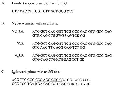
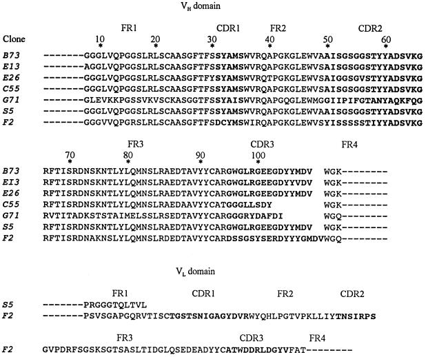
A single-chain Fv (scFv) fusion phage library derived from random combinations of VH and VL (variable heavy and light chains) domains in the antibody repertoire of a vaccinated melanoma patient was previously used to isolate clones that bind specifically to melanoma cells. An unexpected finding was that one of the clones encoded a truncated scFv molecule with most of the VL domain deleted, indicating that a VH domain alone can exhibit tumor-specific binding. In this report a VH fusion phage library containing VH domains unassociated with VL domains was compared with a scFv fusion phage library as a source of melanoma-specific clones; both libraries contained the same VH domains from the vaccinated melanoma patient. The results demonstrate that the clones can be isolated from both libraries, and that both libraries should be used to optimize the chance of isolating clones binding to different epitopes. Although this strategy has been tested only for melanoma, it is also applicable to other cancers. Because of their small size, human origin and specificity for cell surface tumor antigens, the VH and scFv molecules have significant advantages as tumor-targeting molecules for diagnostic and therapeutic procedures and can also serve as probes for identifying the cognate tumor antigens.


Figures

Figure 1
Figure 1
PCR primers for constructing a VH fusion phage library. The direction of the primer sequences is 5′ to 3′. Forward primers are complementary to the sense strand, and back primers are complementary to the antisense strand. The symbols for degenerate nucleotides are as follows: Y = C or T; R = A or G; W = A or T; S = C or G; K = T or G; M = A or C. The SfiI sites are underlined,
Figure 2
Figure 2
Immunohistochemical tests for binding of VH fusion phage C55 and control phage fUSE5 to cultured cells (AH) and frozen tissue sections (IP) as follows. The detection procedure for all panels involved a peroxidase-labeled antiphage second antibody (Pharmacia) followed by the substrate VIP (Vector Laboratories). The slides were counterstained with methyl green and photographed in both brightfield and darkfield. (A) C55/melanoma/bright. (B) C55/melanoma/dark. (C) fUSE5/melanoma/bright. (D) fUSE5/melanoma/dark. (E) C55/melanoma/bright. (F) C55/melanoma/dark. (G) C55/prostate carcinoma/bright. (H) C55/melanocyte/dark. (I) C55/melanoma/bright. (J) C55/melanoma/dark. (K) fUSE5/melanoma/bright. (L) fUSE5/melanoma/dark. (M) C55/normal skin/bright. (N) C55/normal skin/dark. (O) C55/prostate carcinoma/bright. (P) C55/prostate carcinoma/dark. The VIP substrate produces a purple color in brightfield (see A and I) and a golden color in darkfield (see B and J). In I and J the melanoma tissue is marked as m and the connective tissue is marked as c. The melanoma cell line was A2080 and the prostate cell line was DU-145. All of the fusion phage clones shown in Fig. 3 were tested immunohistochemically for binding to the entire panel of cells and sections listed in Table 2. The results showed strong binding to the melanoma cell lines and sections, similar to A, B, I, and J, and no detectable binding to any of the other cells or sections, similar to E, F, G, H, M, N, O, and P.
Figure 3
Figure 3
Amino acid sequences encoded by cDNA inserts in melanoma-specific clones from VH and scFv fusion phage libraries. Clones B73, E13, C55, and G71 were isolated from the VH fusion phage library, and clones S5 and F2 were from the scFv fusion phage library. The amino acid sequences were deduced from the complete nucleotide sequences of the cDNA inserts in the clones; the 15-residue linker between the VH and VL domains of clones S5 and F2 is not shown. The complementarity determining regions (CDRs) are demarcated by boldface type from the framework regions (FRs). The dashed lines at the amino end of the FR1 region and at the carboxyl end of the FR4 regions indicate residues derived from the primers used in the PCR synthesis, The reference positions of the residues are indicated by an asterisk, according to the system of Kabat et al. (4); multiple residues occur at positions 52 (52a) and 82 (82a–c); positions after 100 are designated 100A–K.

References

    1. Cai X, Garen A. Proc Natl Acad Sci USA. 1995;92:6537–6542. - PMC - PubMed
    1. Cai X, Garen A. Proc Natl Acad Sci USA. 1996;93:6280–6285. - PMC - PubMed
    1. Smith P G, Scott J K. Methods Enzymol. 1993;217:228–257. - PubMed
    1. Kabat E A, Wu T T, Reid-Miller M, Perry H M, Gottesman K S. Sequences of Proteins of Immunological Interest. U.S. Government Printing Office, Washington, DC: U.S. Department of Health and Human Services; 1987.
    1. Giudicelli V, Chaume D, Bodmer J, Müller W, Busin C, Marsh S, Bontrop R, Marc L, Malik A, Lefranc M-P. Nucleic Acids Res. 1997;25:206–211. - PMC - PubMed

Publication types

MeSH terms