. 1997 Aug 19;94(17):9320-5.
doi: 10.1073/pnas.94.17.9320.

Targeted overexpression of protein kinase C beta2 isoform in myocardium causes cardiomyopathy

Affiliations

Targeted overexpression of protein kinase C beta2 isoform in myocardium causes cardiomyopathy

H Wakasaki et al. Proc Natl Acad Sci U S A. .

Abstract

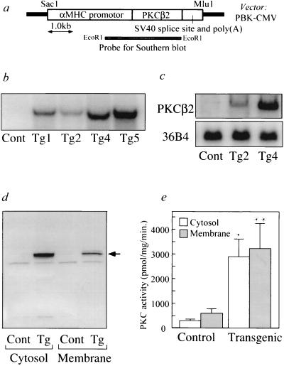
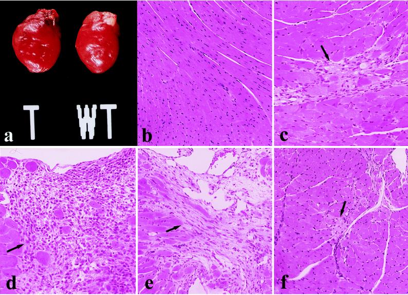
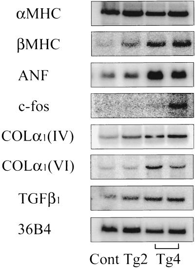
Increased cardiovascular mortality occurs in diabetic patients with or without coronary artery disease and is attributed to the presence of diabetic cardiomyopathy. One potential mechanism is hyperglycemia that has been reported to activate protein kinase C (PKC), preferentially the beta isoform, which has been associated with the development of micro- and macrovascular pathologies in diabetes mellitus. To establish that the activation of the PKCbeta isoform can cause cardiac dysfunctions, we have established lines of transgenic mice with the specific overexpression of PKCbeta2 isoform in the myocardium. These mice overexpressed the PKCbeta2 isoform transgene by 2- to 10-fold as measured by mRNA, and proteins exhibited left ventricular hypertrophy, cardiac myocyte necrosis, multifocal fibrosis, and decreased left ventricular performance without vascular lesions. The severity of the phenotypes exhibited gene dose-dependence. Up-regulation of mRNAs for fetal type myosin heavy chain, atrial natriuretic factor, c-fos, transforming growth factor, and collagens was also observed. Moreover, treatment with a PKCbeta-specific inhibitor resulted in functional and histological improvement. These findings have firmly established that the activation of the PKCbeta2 isoform can cause specific cardiac cellular and functional changes leading to cardiomyopathy of diabetic or nondiabetic etiology.


Figures

Figure 1
Figure 1
(a) Schematic representation of the PKCβ2 isoform transgene. The αMHC promotor includes 5′ regulatory element from rat αMHC gene and heterologous splice cassette. Simian virus 40 (SV40) sequence contains 3′ splicing signal and polyadenylation signal. Restriction enzymes used for generating fragments were as indicated. (b) Southern blot analysis of DNA from heterozygous transgenic mice (Tg1, Tg2, Tg4, and Tg5) and wild-type control (Cont). EcoRI digested mouse tail genomic DNA (15 μg) was hybridized with αMHC–PKCβ2 transgene-specific probe. Size of the positive band is 3.1kb. (c) Northern blot analysis of total RNA (15 μg) isolated from the heart of control (Cont), and transgenic mice (Tg2 and Tg4) hybridized with BamHI fragment of PKCβ2 cDNA. RNA loading differences were normalized using a control cDNA probe (36B4). (d) Immunoblot analysis of cytosolic and membrane fractions isolated from heart of control (Cont) and transgenic mice Tg4 (Tg) using antibodies to PKCβ2 isoform. Arrow indicates the position of endogenous PKCβ2 isoform corresponding to 80 kDa as estimated by prestained protein molecular weight marker (Bio-Rad). (e) PKC activity measurements in cytosolic and membrane fractions from hearts of control (Cont) and transgenic mice Tg4 (Transgenic). Results are the average of four separate experiments. ∗, P < 0.001; ∗∗, P < 0.005 versus nontransgenic control (Student t test).
Figure 2
Figure 2
Cardiac pathology in transgenic mice overexpressing PKCβ2 isoform at 11 weeks of age. (a) Gross photograph demonstrating global enlargement of typical transgenic mouse heart (T) compared with non transgenic littermate (WT). (b) Normal cardiac histology from nontransgenic control mouse. (ce) Photomicrographs of myocardium in PKCβ2 transgenic mouse. (arrows). Microfocal evolving replacement fibrosis, indicative of healing myocyte necrosis (c). Larger epicardial focus of healing with mixed mononuclear inflammatory infiltrate and sharp borders (d). Transmural healing with wall thinning (e). (f) Representative histology of the heart in transgenic mouse treated with PKCβ inhibitor showing marked reduction in size and activity of myocardial lesions (arrow). All panels were stained with hematoxylin and eosin. (b, e, and f, ×120; c and d, ×150.)
Figure 3
Figure 3
Expression of mRNAs for MHC isoforms (αMHC and βMHC), ANF, c-fos, collagen types IV and VI [COLα1 (IV, VI)], and TGFβ1 in PKCβ2 transgenic mice. Northern blot analysis of total heart RNA (15 μg) from control (Cont), transgenic mice (Tg2 and Tg4) at 8–12 weeks of age was performed. RNA loading differences were normalized using a control cDNA probe (36B4) (14).

References

    1. Pucéat M, Brown J H. In: Protein Kinase C. Kuo J F, editor. Oxford: Oxford Univ. Press; 1994. pp. 249–268.
    1. Nishizuka Y. Science. 1992;258:607–614. - PubMed
    1. Strasser R H, Briem S K, Vahl C F, Lang R, Hagl S, Kübler W. Circulation. 1996;94:I551. (abstr.).
    1. Inoguchi T, Battan R, Handler E, Sportsman J R, Heath W, King G L. Proc Natl Acad Sci USA. 1992;89:11059–11063. - PMC - PubMed
    1. Ishii H, Jirousek M R, Koya D, Takagi T, Xia P, Clermont A, Bursell S E, Kern T S, Ballas L M, Heath W F, Stramm L E, Feener E P, King G L. Science. 1996;272:728–731. - PubMed

Publication types

Substances