

Hodgkin and Reed-Sternberg cells in lymphocyte predominant Hodgkin disease represent clonal populations of germinal center-derived tumor B cells
- PMID: 9256483
- PMCID: PMC23186
- DOI: 10.1073/pnas.94.17.9337
Hodgkin and Reed-Sternberg cells in lymphocyte predominant Hodgkin disease represent clonal populations of germinal center-derived tumor B cells
Erratum in
- Proc Natl Acad Sci U S A 1997 Dec 9;94(25):14211
Abstract
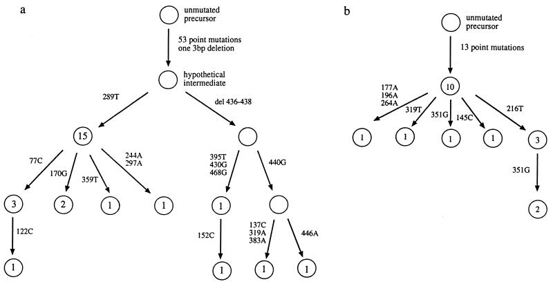
Among the four subtypes of Hodgkin disease (HD), lymphocyte-predominant (LP) HD is now generally considered as a separate entity. The B cell nature of the typical Hodgkin and Reed-Sternberg (HRS) cells and their variants (L and H, lymphocytic and histiocytic cells) in LP HD has long been suspected, but the question of whether these cells represent a true tumor clone is unclear. We previously demonstrated clonal Ig gene rearrangements in one case of LP HD. In the present study, five cases of LP HD were analyzed by micromanipulation of single HRS cells from frozen tissue sections and DNA amplification of rearranged Ig heavy chain genes from those cells. Clonal V gene rearrangements harboring somatic mutations were detected in each case. In three cases ongoing somatic mutation was evident. This shows that HRS cells in LP HD are a clonal tumor population derived from germinal center B cells. The pattern of somatic mutation indicates that HRS cells in LP HD are selected for antibody expression. This, and the presence of ongoing mutation discriminates LP from classical HD.
Figures
References
-
- Burke J S. In: Neoplastic Hematopathology. Knowles D M, editor. Baltimore: Williams & Wilkins; 1992. pp. 497–533.
-
- Harris N L, Jaffe E S, Stein H, Banks P M, Chan J K C, Cleary M L, Delsol G, de Wolf-Peeters C, Falini B, Gatter K C, Grogan T M, Isaacson P G, Knowles D M, Mason D Y, Müller-Hermelink H K, Pileri S A, Piris M A, Rafkiaer E, Warnke R A. Blood. 1994;84:1361–1392. - PubMed
-
- Mason D Y, Banks P M, Chan J, Cleary M L, Delsol G, de Wolf-Peeters C, Falini B, Gatter K, Grogan T M, Harris N L, Isaacson P G, Jaffe E S, Knowles D M, Müller-Hermelink K, Pileri S, Ralfkiaer E, Stein H, Warnke R. Am J Surg Pathol. 1994;18:526–530. - PubMed
-
- Klein U, Küppers R, Rajewsky K. Eur J Immunol. 1993;23:3272–3277. - PubMed
Publication types
MeSH terms
Substances
Associated data
- Actions
- Actions
- Actions
- Actions
- Actions
LinkOut - more resources
Full Text Sources
Other Literature Sources
Medical
