. 1997 Aug 19;94(17):9400-5.
doi: 10.1073/pnas.94.17.9400.

Construction of hybrid proteins that migrate retrogradely and transynaptically into the central nervous system

Affiliations

Construction of hybrid proteins that migrate retrogradely and transynaptically into the central nervous system

L Coen et al. Proc Natl Acad Sci U S A. .

Abstract

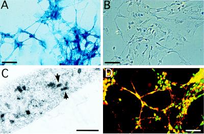
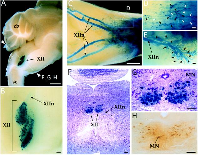
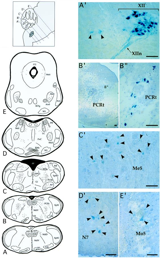
The nontoxic proteolytic C fragment of tetanus toxin (TTC peptide) has the same ability to bind nerve cells and be retrogradely transported through a synapse as the native toxin. We have investigated its potential use as an in vivo neurotropic carrier. In this work we show that a hybrid protein encoded by the lacZ-TTC gene fusion retains the biological functions of both proteins in vivo-i.e. , retrograde transynaptic transport of the TTC fragment and beta-galactosidase enzymatic activity. After intramuscular injection, enzymatic activity could be detected in motoneurons and connected neurons of the brainstem areas. This strategy could be used to deliver a biological activity to neurons from the periphery to the central nervous system. Such a hybrid protein could also be used to map synaptic connections between neural cells.


Figures

Figure 1
Figure 1
In vitro uptake of β-gal–TTC hybrid protein and control β-gal by 1009 neural cells. Eight days after the start of retinoic treatment, differentiated 1009 cells were incubated at 37°C for 2 hr with 5 μg/ml of each protein and fixed. X-Gal staining was performed after incubation with β-gal–TTC (A) or β-gal (B). Strong labeling was specifically detected in neural cells. (C) Electron micrograph of X-Gal-stained neural cell axon showing intracellular localization of the fusion protein: arrows indicate the precipitate probably associated with filaments. After β-gal–TTC incubation, immunodetection was performed with anti-neurofilament and anti-TTC antibodies (D): colabeling showing that β-gal–TTC was only uptaken in neuronal cells. [Bars = 100 μm (A, B, and D) and 1 μm (C).]
Figure 2
Figure 2
Retrograde labeling following β-gal–TTC intramuscular injection in the tongue of mice. A total of 100 μg of protein was injected, and observations were made 12 hr (A, B, C, D, and E) and 18 hr (F, G, and H) postinjection. In toto X-Gal staining on brain clearly showed the hypoglossal structure (XII) with its two nuclei retrogradely labeled (A and B). The hypoglossal nerve (XIIn) was also intensively stained (C), as was its arborization (E, see arrows). Putative neuromuscular junctions (D) with button-like structure (black arrows) and the terminal arborization of axons (white arrows) are visible. (F and G with different magnification) Histological analysis of brain slices after X-Gal staining. The hybrid protein is localized in XII motoneurons cytoplasm. (H) Immunodetection with anti-TTC antibody showing a colocalization with the β-gal activity. [Bars = 1 mm (A and C), 100 μm (B and DH).] cb, Cerebellum; sc, spinal cord; MN, motoneuron.
Figure 3
Figure 3
Transneuronal labeling following intramuscular injection of β-gal–TTC into the mouse tongue. A total of 100 μg of protein was injected, and observations were made 24–48 hr postinjection. (AE) Distribution of β-gal-positive neurons in different brainstem sections, also summarized in Table 1. One dot represents one labeled neuron; six animals were analyzed. (A′–E′) Examples of labeled neurons: labeling in medullary reticular dorsal (MdD) area (A′), arrows showing second order stained neurons; labeling in parvicellular reticular nucleus (PCRt) area (B′ and B" with different magnification) showing a β-gal-positive column of neurons; labeling in Me5 area (C′), arrows show second-order stained neurons; labeling in N7 (D′) and Mo5 areas (E′), arrows show β-gal-positive neurons. [Bars = 100 μm (A′, B′, B", D′, and E′) and 50 μm (C′).] AP, area postrema; Aq, Aqueduct (Sylvius); 4V, 4th ventricule; Gi, gigantocellular reticular nucleus (nu); IO, inferior olive; KF, Kölliker–Fuse nu; LRt, lateral reticular nu; PB, parabrachial nu; Pr5, principal sensory trigeminal nu; RtTg, reticulotegmental nu pons; SO, superior olive; Sp5C, spinal trigeminal nu, caudal part; Sp5I, spinal trigeminal nu, interpolar part; Sp5O, spinal trigeminal nu, oral part; see Table 1 for other abbreviations.

References

    1. Eisel U, Jarausch W, Goretzki K, Henschen A, Engels J, Weller U, Hudel M, Habermann E, Niemann H. EMBO J. 1986;5:2495–2502. - PMC - PubMed
    1. Fairweather N F, Lyness V A. Nucleic Acids Res. 1986;14:7809–7812. - PMC - PubMed
    1. Montecucco C, Schiavo G. Q Rev Biophys. 1995;28:423–472. - PubMed
    1. Schwab M E, Thoenen H. Brain Res. 1976;105:213–227. - PubMed
    1. Schwab M E, Thoenen H. Brain Res. 1977;122:459–474. - PubMed

Publication types

LinkOut - more resources