. 1997 Aug 19;94(17):9422-7.
doi: 10.1073/pnas.94.17.9422.

A single dose of kainic acid elevates the levels of enkephalins and activator protein-1 transcription factors in the hippocampus for up to 1 year

Affiliations

A single dose of kainic acid elevates the levels of enkephalins and activator protein-1 transcription factors in the hippocampus for up to 1 year

G Bing et al. Proc Natl Acad Sci U S A. .

Abstract

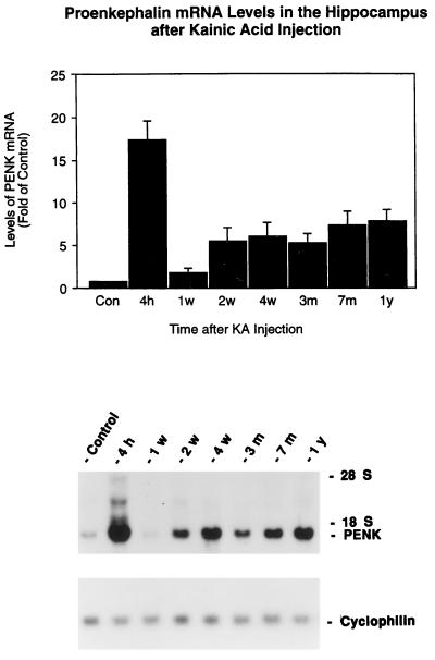
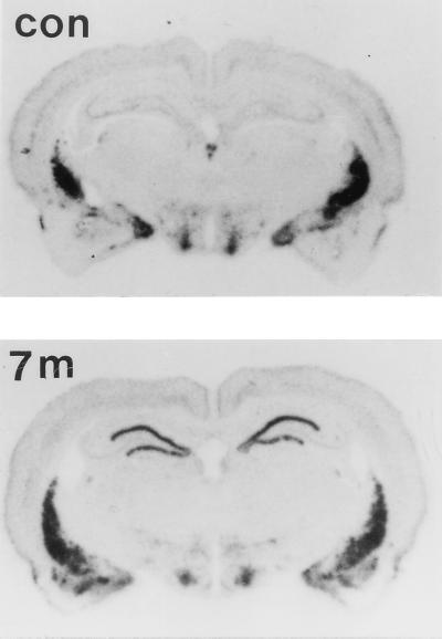
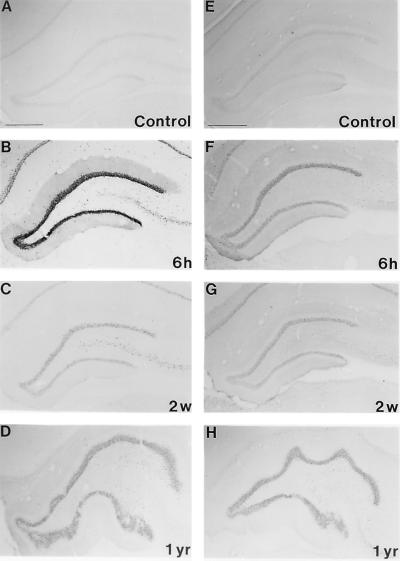
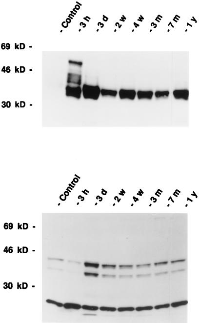
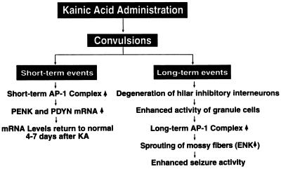
Neuronal plasticity plays a very important role in brain adaptations to environmental stimuli, disease, and aging processes. The kainic acid model of temporal lobe epilepsy was used to study the long-term anatomical and biochemical changes in the hippocampus after seizures. Using Northern blot analysis, immunocytochemistry, and Western blot analysis, we have found a long-term elevation of the proconvulsive opioid peptide, enkephalin, in the rat hippocampus. We have also demonstrated that an activator protein-1 transcription factor, the 35-kDa fos-related antigen, can be induced and elevated for at least 1 year after kainate treatment. This study demonstrated that a single systemic injection of kainate produces almost permanent increases in the enkephalin and an activator protein-1 transcription factor, the 35-kDa fos-related antigen, in the rat hippocampus, and it is likely that these two events are closely associated with the molecular mechanisms of induction of long-lasting enhanced seizure susceptibility in the kainate-induced seizure model. The long-term expression of the proenkephalin mRNA and its peptides in the kainate-treated rat hippocampus also suggests an important role in the recurrent seizures of temporal lobe epilepsy.


Figures

Figure 1
Figure 1
Time course of the expression of PENK mRNA in the rat hippocampus after kainate injection. Rats were injected with kainate (7.25 mg/kg) s.c.. Hippocampi (n = 4) were collected at different time points and analyzed with Northern blots. (Upper) The autoradiographic signals corresponding to PENK and cyclophilin mRNA were quantified by laser densitometry. Ratios of PENK/cyclophilin mRNA density were calculated. Data represent means ± SEM. P < 0.01 at all time points compared with control except at 1 week (two-way analysis of variance, Bonferroni–Dunn post hoc tests were performed for between groups comparison). Control represents the mean of different time points from 1 week, 3 months, 7 months, and 1 year after a single injection of saline. There was no significant difference in the levels of PENK mRNA from these of control hippocampi. (Lower) Representative autoradiogragh of a Northern blot probed with 32P-labeled PENK and cyclophilin cRNAs showing the effects at 4 hr, 1 week, 2 weeks, 1 month, 3 months, 7 months, and 1 year after kainate treatment. The control sample was from the 3-month time point.
Figure 2
Figure 2
Autoradiographs of in situ hybridization for PENK mRNA on coronal sections of the rat brain. Representative autoradiographs from four independent experiments were taken from a control animal 7 months after saline injection and 7 months after kainate treatment. Autoradiographs were made from x-ray film 7 days after exposure to brain sections hybridized with 35S-labeled probes.
Figure 3
Figure 3
Expression of ENK-IR in the rat hippocampus after kainate treatment. Representative immunocytochemical photomicrographs from three separate experiments are shown. Hippocampal immunostainings for ENK-IR from saline-treated rats at different time points were similar; photomicrographs from 6 hr (6 h) and 1 year (1 y) are shown. The kainate-treated groups include 6 hr (6 h), 2 weeks (2 w), 3 weeks (3 w), 1 month (1 m), 3 months (3 m), 7 months (7 m), and 1 year (1 y). Dramatic increases in ENK-IR were observed at 3 weeks after kainate treatment and persisted for up to 1 year. Note the progressive increase in the density of ENK-IR in the inner molecular layer of the dentate gyrus (sprouting of mossy fibers) in kainate-treated animals. (Scale bar = 400 μm.)
Figure 4
Figure 4
Representative photomicrographs from three separate experiments of FRA-like (AD) and JunD-like (EH) immunoreactivity in the hippocampus. The immunostained sections are from rats 6 hr (6 h), 2 weeks (2 w), and 1 year (1 yr) after treatment with kainate. Immunostaining for control from a 2-week (Control) time point is shown. There are no significant differences in the levels of FRA and JunD immunoreactivities between hippocampi 6 hr, 2 weeks, and 1 year after saline injection. Note the long-lasting changes in the intensity of the staining.
Figure 5
Figure 5
Time course of the expression of FRA-like (Upper) and JunD-like (Lower) immunoreactivity after kainate administration as revealed by Western blot analysis. Representative photomicrographs from four separate experiments are shown. Immunostaining for a control rat from a 3-month (Control) time point is shown. There were no significant differences in the levels of FRA and JunD immunoreactivity from the control hippocampi at different time points. The kainate-injected groups were sacrificed after 3 hr (3 h), 3 days (3 d), 2 weeks (2 w), 4 weeks (4 w), 3 months (3 m), 7 months (7 m), and 1 year (1 y). Note that at least four bands of protein were recognized by the FRA antiserum at 3 hr after kainate treatment, but only one 35-kDa FRA was detected at 2 weeks after kainate treatment. JunD antiserum appears to recognize three bands of immunoreactivity at 43, 39, and 28 kDa.
Figure 6
Figure 6
Flow diagram of the biochemical and morphological changes in the hippocampus after treatment with kainate. Single systemic injections of kainate result in both short-term and long-term changes in the hippocampus. The short-term event is reflected by the rapid induction of AP-1 transcription factors, which may underlie the rapid induction of PENK and PDYN in the hippocampus. Elevated expression of the opioid peptides in the hippocampus lasted for a few days and returned to normal levels. This is the end of the short-term event. The long-term event after the kainate injection may ultimately lead to increases in the seizure susceptibility of the animals. Degeneration of the hilar inhibitory interneurons is the key event, which sets up the following events. Disinhibition of dentate granule cells increases changes of granule cell activity. This may activate the long-term AP-1 transcription factor. The loss of the targets of the mossy fibers will result in sprouting. These may induce the second phase of the increase of PENK expression in the granule cells. All of these events will result in increases of seizure activities.

References

    1. Sloviter R S, Dempster D W. Brain Res Bull. 1985;15:39–60. - PubMed
    1. Represa A, Ben-Ari Y. Epilepsy Res Suppl. 1992;7:261–269. - PubMed
    1. Tauck D L, Nadler J V. J Neurosci. 1985;5:1016–1022. - PMC - PubMed
    1. Nadler J V, Okazaki M M, Gruenthal M, Ault B, Armstrong D R. Adv Exp Med Biol. 1986;203:673–686. - PubMed
    1. Sutula T, Cavazos J, Golarai G. J Neurosci. 1992;12:4173–4187. - PMC - PubMed