. 1997 Aug 19;94(17):9445-50.
doi: 10.1073/pnas.94.17.9445.

Essential role of POU-domain factor Brn-3c in auditory and vestibular hair cell development

Affiliations

Essential role of POU-domain factor Brn-3c in auditory and vestibular hair cell development

M Xiang et al. Proc Natl Acad Sci U S A. .

Abstract

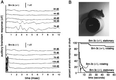
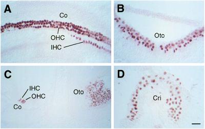
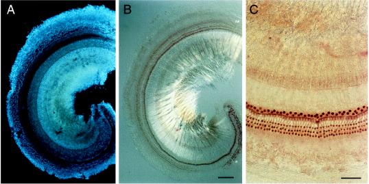
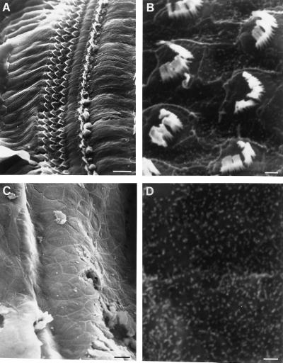
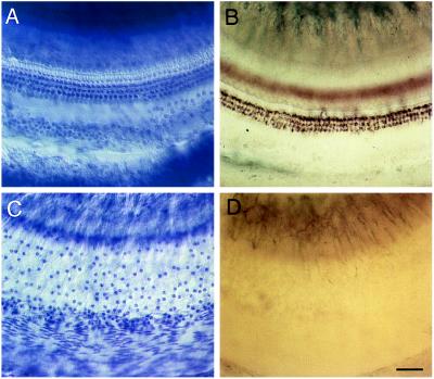
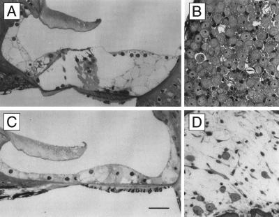
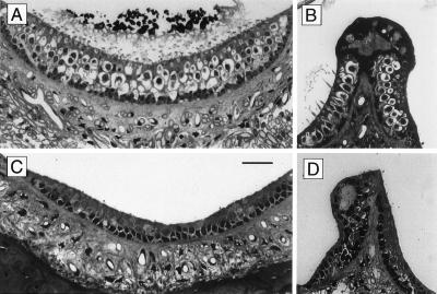
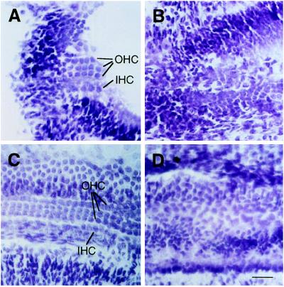
The Brn-3 subfamily of POU-domain transcription factor genes consists of three highly homologous members-Brn-3a, Brn-3b, and Brn-3c-that are expressed in sensory neurons and in a small number of brainstem nuclei. This paper describes the role of Brn-3c in auditory and vestibular system development. In the inner ear, the Brn-3c protein is found only in auditory and vestibular hair cells, and the Brn-3a and Brn-3b proteins are found only in subsets of spiral and vestibular ganglion neurons. Mice carrying a targeted deletion of the Brn-3c gene are deaf and have impaired balance. These defects reflect a complete loss of auditory and vestibular hair cells during the late embryonic and early postnatal period and a secondary loss of spiral and vestibular ganglion neurons. Together with earlier work demonstrating a loss of trigeminal ganglion neurons and retinal ganglion cells in mice carrying targeted disruptions in the Brn-3a and Brn-3b genes, respectively, the Brn-3c phenotype reported here demonstrates that each of the Brn-3 genes plays distinctive roles in the somatosensory, visual, and auditory/vestibular systems.


Figures

Figure 1
Figure 1
Auditory brainstem responses and balancing defects in Brn-3c (−/−) mice. (A) Representative auditory brainstem responses to a click stimulus are shown at different stimulus intensities. Brn-3c (+/−) mice show a threshold between 49 and 59 dB SPL and robust responses at and above 59 dB SPL. Brn-3c (−/−) mice show no response at any stimulus level, including 109 dB SPL, the highest level shown here. The traces at 109 dB SPL show a stimulus artifact in the first 1 μsec. Traces were produced by averaging over 1,000 responses. (B Upper) A Brn-3c (+/−) mouse is shown balancing on a stationary drum. (Lower) Percentage of animals remaining on the drum during the 60 sec following their placement on it. Each of seven animals of the indicated genotypes was tested in 10 trials in which the drum was stationary and in 10 trials in which the drum was rotating at 7 rpm, a total of 70 trials for each genotype and test condition.
Figure 2
Figure 2
Brn-3c in the developing mouse inner ear. Immunostaining with anti-Brn-3c antibodies of frozen sections at E17.5 (A and B) and E19.5 (C and D). Staining is present in the nuclei of developing hair cells at all stages shown, and is absent from other cell types. Cri, crista; Co, cochlea; Oto, otolith organ; IHC, inner hair cells; OHC, outer hair cells. [Scale bar = 50 μm (C) and 25 μm (A, B, and D).]
Figure 3
Figure 3
Brn-3c in the adult organ of Corti. One turn of a wild-type organ of Corti adjacent to the apex is shown. The cochlea was immunostained with affinity-purified anti-Brn-3c antibodies and the dissected organ of Corti was then incubated with DAPI. (A) DAPI staining reveals nuclei of supporting cells that are not immunostained. (B and C) Anti-Brn3c immunoreactivity is found exclusively within the single row of inner hair cell nuclei and the three rows of outer hair cell nuclei. All hair cell nuclei are immunostained. Note that the precipitate formed by reaction of 3-amino-9-ethylcarbozole, the immunoperoxidase substrate, quenches DAPI fluorescence. [Scale bar = 100 μm (A and B) and 50 μm (C).]
Figure 4
Figure 4
Scanning electron microscopy of the organ of Corti in Brn-3c (+/−) and (−/−) mice. (A and B) The apical surface of the organ of Corti in Brn-3c (+/−) mice shows the wild-type pattern of stereociliary bundles. (A) Stereociliary bundles emanating from the three rows of outer hair cells are seen to the left of the stereociliary bundles emanating from a single row of inner hair cells. The four rows of hair cells are flanked by supporting cells. (B) A different region of the organ of Corti at high magnification shows stereociliary bundles from two rows of outer hair cells protruding above the microvilli that cover the apical face of the supporting cells. (C and D) The apical surface of the organ of Corti in Brn-3c (−/−) mice lacks stereociliary bundles. (D) At high magnification the apical surface is seen to be composed of cells bearing microvilli. [Scale bars = 10 μm (A and C) and 1 μm (B and D).]
Figure 5
Figure 5
Whole-mount preparation of the organ of Corti in Brn-3c (+/+), (+/−), and (−/−) mice. (A) Organ of Corti from a Brn-3c (+/−) mouse stained with cresyl violet shows the wild-type arrangement of a single row of inner hair cells (out of the focal plan) and three rows of outer hair cells (in the focal plane). (B) Organ of Corti from a Brn-3c (+/+) mouse stained histochemically for acetylcholine esterase reveals efferent synapses on the three rows of outer hair cells. By contrast, the organ of Corti from a Brn-3c (−/−) mouse lacks identifiable hair cells (C) and cholinergic innervation (D). (Scale bar = 50 μm.)
Figure 6
Figure 6
Absence of hair cells in the organ of Corti and defects in the spiral ganglion in Brn3c (−/−) mice. (A) Organ of Corti from a Brn-3c (+/−) mouse. The single inner hair cell, the three outer hair cells, and the three underlying Deiter’s cells are clearly visible. (B) Part of a spiral ganglion from a Brn-3c (+/−) mouse showing a dense packing of myelinated axons and neuronal cell bodies. (C) Organ of Corti from a Brn-3c (−/−) mouse. Inner and outer hair cells are missing and the epithelium beneath the tectorial membrane contains only supporting cells. (D) The spiral ganglion from a Brn-3c (−/−) mouse contains fewer than one-tenth as many myelinated axons and neuronal cell bodies than the spiral ganglia of Brn-3c (+/+) or (+/−) mice. Tissues were embedded in Spurr’s resin and 1 μm sections were stained with methylene blue. (Scale bar = 25 μm.)
Figure 7
Figure 7
Absence of hair cells in the otolith organs and cristae of Brn-3c (−/−) mice. (A) An otolith organ from a Brn-3c (+/−) mouse. Type I hair cells, the most abundant hair cell class, have a lightly stained nerve chalice surrounding a darkly stained cell body. Ciliary bundles can be seen protruding into the densely stained otoliths at the top. A single layer of supporting cells lies beneath the layer of hair cells. (B) Crista from a Brn-3c (+/+) mouse. Type I hair cells are abundant and their ciliary bundles are seen at the left side of the ampullary crest, where the section is nearly perpendicular to the apical surface. (C and D) Otolith organ (C) and crista (D) from a Brn-3c (−/−) mouse are devoid of hair cells. Presumptive supporting cells are seen within the epithelium. The density of axon bundles beneath the otolith organ is greatly reduced in the Brn-3c (−/−) animal. Tissues were embedded in Spurr’s resin and 1 μm sections were stained with methylene blue. (Scale bar = 25 μm.)
Figure 8
Figure 8
Defects during development of the cochlea of Brn-3c (−/−) mice. Cresyl violet-stained frozen sections showing the progressive development of the organ of Corti at P1 (A and B) and P5 (C and D). (A and C) Brn-3c (+/−). (B and D) Brn-3c (−/−). The organized layering of inner and outer hair cells fails to appear in Brn-3c (−/−) animals. HC, hair cells; IHC, inner hair cells; OHC, outer hair cells. (Scale bar = 25 μm.)

References

    1. Jan Y N, Jan L. Cell. 1993;75:827–830. - PubMed
    1. Lee J E, Hollenberg S M, Snider L, Turner D L, Lipnick N, Weintraub H. Science. 1995;268:836–844. - PubMed
    1. Keynes R, Krumlauf R. Annu Rev Neurosci. 1994;17:109–132. - PubMed
    1. Ryan A K, Rosenfeld M G. Genes Dev. 1997;11:1207–1225. - PubMed
    1. Herr W, Sturm R A, Clerc R G, Corcoran L M, Baltimore D, Sharp P A, Ingraham H A, Rosenfeld M G, Finney M, Ruvkin G, Horvitz H R. Genes Dev. 1988;2:1513–1516. - PubMed

Publication types