. 1997 Aug 19;94(17):9493-8.
doi: 10.1073/pnas.94.17.9493.

Myofibrillogenesis visualized in living embryonic cardiomyocytes

Affiliations

Myofibrillogenesis visualized in living embryonic cardiomyocytes

G A Dabiri et al. Proc Natl Acad Sci U S A. .

Abstract

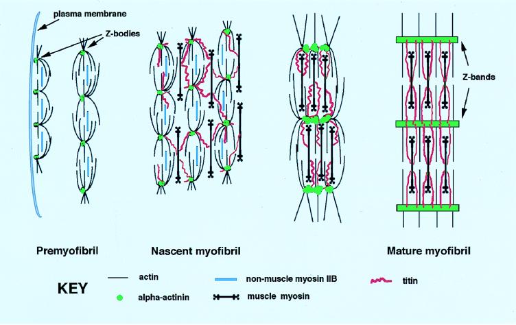
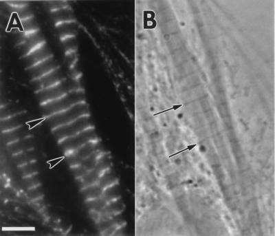
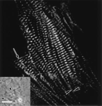
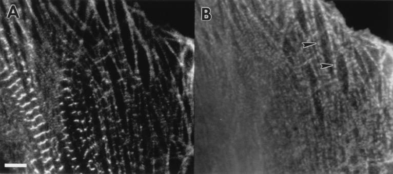
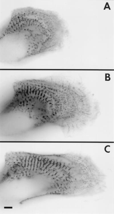
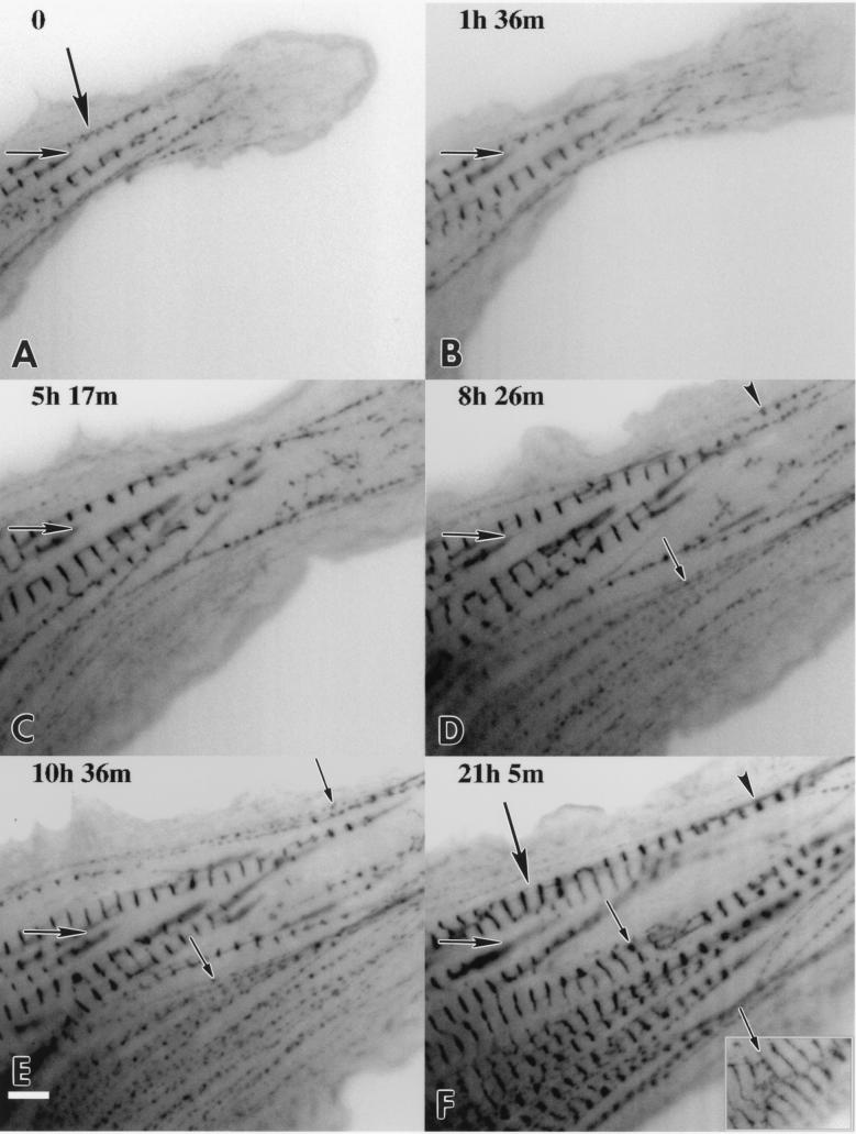
Myofibril formation was visualized in cultured live cardiomyocytes that were transfected with plasmids expressing green fluorescent protein (GFP) linked to the Z-band protein, alpha-actinin. The expression of this fluorescent protein provided an in vivo label for structures containing alpha-actinin. The GFP-alpha-actinin fusion protein was incorporated into Z-bands, intercalated discs, and attachment plaques, as well as into the punctate aggregates, or Z-bodies, that are thought to be the precursors of Z-bands. Observations of live cells over several days in culture permitted us to test aspects of several theories of myofibril assembly that had been proposed previously based on the study of fixed cells. Fine fibrils, called premyofibrils, that formed de novo at the spreading edges of cardiomyocytes, contained punctate concentrations of alpha-actinin, termed Z-bodies. The punctate Z-bodies grew and aligned with Z-bodies in adjacent fibrils. With increasing time, adjacent fibrils and Z-bodies appeared to fuse and form mature myofibrils and Z-bands in cytoplasmic regions where the linear arrays of Z-bodies had been. These new myofibrils became aligned with existing myofibrils at their Z-bands to form myofibrils that spanned the length of the spread cell. These results are consistent with a model that postulates that the fibrils that form de novo near the cell membrane are premyofibrils-i.e., the precursors of mature myofibrils.


Figures

Figure 1
Figure 1
Model for the assembly of a mature myofibril based on the data presented in this and previous papers (1, 2). The position of α-actinin (shaded circles) relative to actin, muscle myosin II, nonmuscle myosin IIB, and titin is shown for each stage of myofibril development. Near the spreading edge of the cell (far left), α-actinin is found in mini-sarcomeric arrays in the premyofibrils. In the nascent myofibril, the α-actinin-rich Z-bodies have begun to laterally associate, possibly through interaction with titin. Overlapping muscle myosin II filaments bound to titin are present at this stage. In the mature myofibril the α-actinin containing Z-bodies fuse to form the wide lateral arrays of mature Z-bands; nonmuscle myosin IIB is lost whereas muscle myosin filaments are now aligned into A-bands. The Z-bodies grow in size in the progression from premyofibril to mature myofibril.
Figure 2
Figure 2
(A) Fluorescent image of an embyronic chicken cardio-myocyte transfected with the α-actinin–GFP plasmid showing the colocalization of α-actinin–GFP (arrowheads) with the phase-dense Z-bands (arrows) (B). (Bar = 5 μm.)
Figure 3
Figure 3
Fluorescent image of an actively contracting embryonic chicken cardiomyocyte transfected with α-actinin–GFP. The fusion protein localizes to the Z-bands of the mature contracting myofibrils, the Z-bodies of the premyofibrils (arrowheads), and an intercalated disc (arrow) that is shown in phase-contrast in the inset (arrow). (Bar = 5 μm.)
Figure 4
Figure 4
Fluorescent images of an embryonic chicken cardiomyocyte (A) transfected with α-actinin and (B) fixed and stained with a nonmuscle myosin IIB antibody. Nonmuscle myosin IIB is in the premyofibrils at the edge of the cardiomyocyte (B, arrowheads) but not in the mature myofibrils in the central region of the cell. (Bar = 5 μm.)
Figure 5
Figure 5
Time-lapse fluorescent images of a spreading and beating embyronic chicken cardiomyocyte transfected with α-actinin–GFP are shown as reverse contrast at the initial time point (A) 0 h, (B) 3 h later, and (C) 10 h. Four focal planes were digitally added for each time point to identify all the Z-bodies. (Bar = 5 μm.)
Figure 6
Figure 6
Time-lapse fluorescent image observation of a spreading and beating embyronic chicken cardiomyocyte transfected with α-actinin–GFP shown in reverse contrast with the time of acquisition relative to A indicated on each micrograph (BF). The same prominent adhesion plaque is marked in each panel as a reference point (AF, horizontal arrows). Note the extent of spreading with respect to this plaque. As the cell spreads toward the lower part of the image, punctate Z-bodies assemble into linear arrays that mature into Z-bands of myofibrils. One myofibril indicated by a large arrow (A) appears to double in length over 21 h. This appears to occur by lateral coalescence of myofibrils (DF, arrowheads). Adjacent Z-bodies (D, small arrow) increase in diameter (E, small arrow) and fuse into a mature Z-band (F, small arrow). At 28 h the Z-bands became thinner and less punctate (F Inset, small arrow). (Bar = 5 μm.)

Similar articles

Cited by

References

    1. Rhee D, Sanger J M, Sanger J W. Cell Motil Cytoskeleton. 1994;28:1–24. - PubMed
    1. Turnacioglu K K, Mittal B, Dabiri G A, Sanger J M, Sanger J W. Cell Struct Funct. 1997;22:73–82. - PubMed
    1. Dlugosz A A, Antin P B, Nachmias V T, Holtzer H. J Cell Biol. 1984;99:2268–2278. - PMC - PubMed
    1. Schultheiss T, Lin Z, Lu M-H, Murray J, Fischman D A, Weber K, Masaki T, Imamura M, Holtzer H. J Cell Biol. 1990;110:1159–1172. - PMC - PubMed
    1. Epstein H F, Fischman D A. Science. 1991;251:1039–1044. - PubMed

Publication types

LinkOut - more resources