. 1997 Aug 19;94(17):9504-9.
doi: 10.1073/pnas.94.17.9504.

Isolation and characterization of an amino acid-selective channel protein present in the chloroplastic outer envelope membrane

Affiliations

Isolation and characterization of an amino acid-selective channel protein present in the chloroplastic outer envelope membrane

K Pohlmeyer et al. Proc Natl Acad Sci U S A. .

Abstract

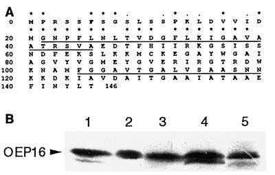
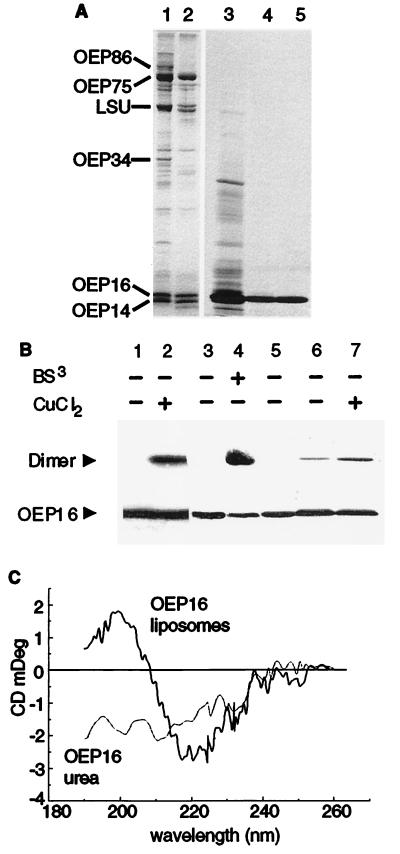
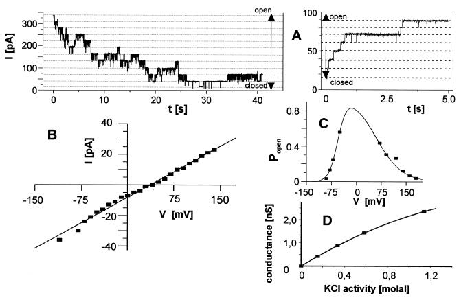
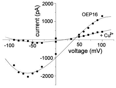
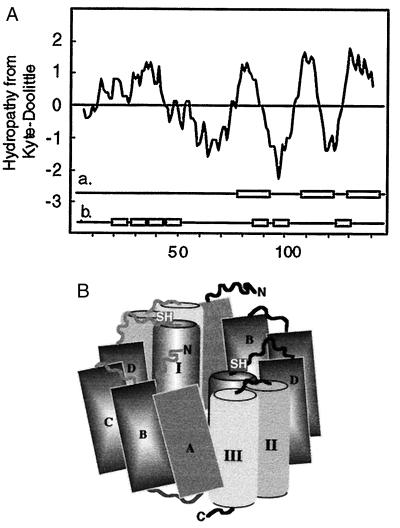
The reconstituted pea chloroplastic outer envelope protein of 16 kDa (OEP16) forms a slightly cation-selective, high-conductance channel with a conductance of Lambda = 1,2 nS (in 1 M KCl). The open probability of OEP16 channel is highest at 0 mV (Popen = 0.8), decreasing exponentially with higher potentials. Transport studies using reconstituted recombinant OEP16 protein show that the OEP16 channel is selective for amino acids but excludes triosephosphates or uncharged sugars. Crosslinking indicates that OEP16 forms a homodimer in the membrane. According to its primary sequence and predicted secondary structure, OEP16 shows neither sequence nor structural homologies to classical porins. The results indicate that the intermembrane space between the two envelope membranes might not be as freely accessible as previously thought.


Figures

Figure 1
Figure 1
Deduced amino acid sequence and tissue distribution of pea OEP16. (A) The deduced amino acid sequence of the pea cDNA clone for OEP16 is shown (accession no. Z73553). The peptide sequences obtained after CNBr cleavage are underlined. N-terminal 40 amino acids of the OEP16 are compared with homologues from arabidopsis, maize, and rice. An asterisk indicates identities, and dots indicate homologous exchanges in all four species, respectively. (B) Immunodetection of OEP16 in chloroplasts from leaves (lane 2), shoots (lane 3), and root-plastids (lane 4) of light-grown pea plants or in leaves from etiolated plants (lane 5). Total plastid protein (100 μg) was used. Lane 1, presence of OEP16 in purified chloroplastic outer envelopes (1 μg protein). Organelles were isolated and subfractionated as described (–35).
Figure 2
Figure 2
Characterization of pea OEP16. (A) Chloroplastic outer membrane polypeptides were analyzed by SDS/PAGE either before (lane 1) or after (lane 2) treatment with the protease thermolysin (24, 25). The position of known polypeptides is indicated. Silver-stained polypeptide composition of heterologuously expressed OEP16 recovered in inclusion bodies (lane 3) after anion- (lane 4) and cation-exchange chromatography (lane 5). (B) OEP16 forms a homodimer. Purified outer envelope membranes (10 μg protein) were either not treated or treated with the crosslinker BS3 (500 μM, 10 min on ice) or with 1 mM CuCl2 (lanes 1–4). E. coli-expressed OEP16 (lane 5) was reconstituted into liposomes and incubated in the absence (lane 6) or presence (lane 7) of 1 mM CuCl2. Products were separated by SDS/PAGE (36) in the absence of reducing agents and analyzed by immunoblotting using αOEP16. (C) CD spectra of OEP16 either denatured in 8 M urea or reconstituted into liposomes.
Figure 3
Figure 3
Reconstituted OEP16 constitutes a voltage-sensitive, cation-selective pore. (A) Liposomes containing E. coli-overexpressed OEP16 were fused to black lipid membranes. The cis chamber contained 250 mM KCl and the trans chamber, 20 mM KCl. A current trace from a bilayer containing 11 active pores is shown directly after a voltage jump from 0 to 150 mV (Left). The channels, which were open at 0 mV, close after the increase to 150 mV. When the identical experiment (Left) was continued, opening of several channels was measured after lowering the potential in one step to 80 mV (Right). (B) Current–voltage relationship from the data presented in A. The slope of the linear regression is 330 pS and the zero current potential is 32 mV. (C) Open probabilities of the OEP16 pore. Conditions were as in A, except that 250 mM KCl was used on both sides of the bilayer. Voltages were applied for 10 min, but only the mean current of the last minute was taken as the steady-state current. The quotient of this current and the total current for 24 open channels (estimated number of channels present in this experiment) are plotted. All potentials are referred to the trans compartment. (D) The conductivity of OEP16 depends nearly linear on solution activity of KCl. All measurements were performed on the same bilayer. Solutions were exchanged by perfusion, and current traces were recorded for at least eight different voltages.
Figure 4
Figure 4
Current–voltage relationship of bilayers containing multiple copies of OEP16 channels in the absence and presence of 10 mM CuCl2 on the cis side (cis compartment 250 mM KCl, trans 20 mM KCl). Mean values were calculated from 5-s current-recording intervals at the given voltages. Considering the single-channel conductance of OEP16 (330 pS in 250 mM KCl), the bilayer contained about 200 active channels.
Figure 5
Figure 5
Protein structure prediction of OEP16. (A) A hydropathy analysis was carried out according to the Kyte–Doolittle algorithm using a window of 11 amino acids. The prediction for the presence of hydrophobic α-helices (a) was done according to ref. . The possible presence of β-strands in OEP16 (b) was predicted according to ref. . (B) Hypothetic model for the membrane arrangement of the pea OEP16 dimer. Putative β-strands A–D are shown as rectangles; the putative helices are shown as columns. The position of cysteine-71 is indicated by an SH. The model is not drawn to scale.

Similar articles

Cited by

References

    1. Gray J C, Row P E. Trends Cell Biol. 1995;5:243–251. - PubMed
    1. Schnell D J. Cell. 1995;83:521–524. - PubMed
    1. Soll J. Bot Acta. 1995;108:277–282.
    1. Joyard J, Block M A, Douce R. Eur J Biochem. 1991;199:489–509. - PubMed
    1. Lam H-M, Coschigano K T, Oliveira I C, Melo-Oliveira R, Coruzzi G M. Annu Rev Plant Physiol Plant Mol Biol. 1996;47:569–593. - PubMed

Publication types

Associated data

LinkOut - more resources