. 1997 Aug 25;138(4):759-69.
doi: 10.1083/jcb.138.4.759.

Acid-induced dissociation of VacA, the Helicobacter pylori vacuolating cytotoxin, reveals its pattern of assembly

Affiliations

Acid-induced dissociation of VacA, the Helicobacter pylori vacuolating cytotoxin, reveals its pattern of assembly

T L Cover et al. J Cell Biol. .

Abstract

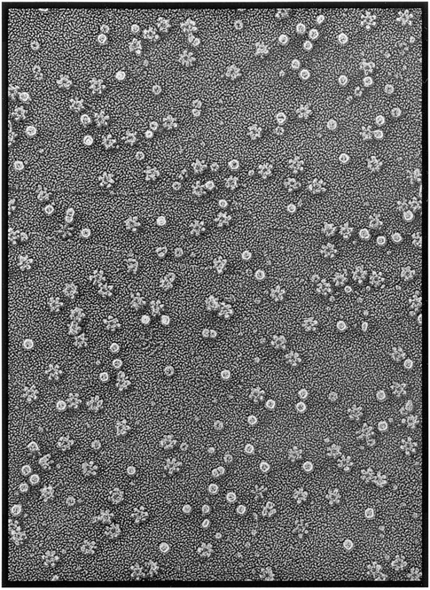
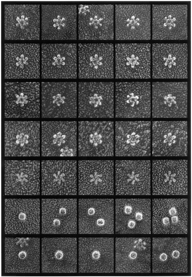
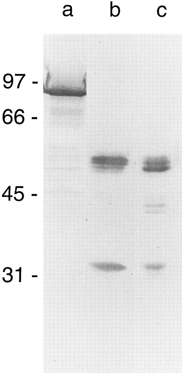
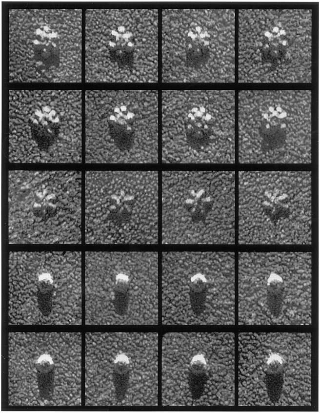
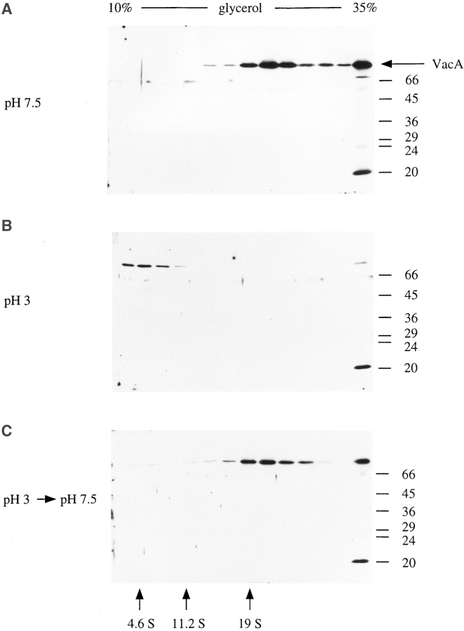
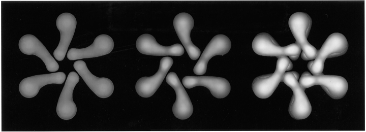
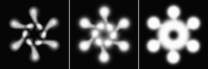
In this study, we describe the ultrastructural changes associated with acid activation of Helicobacter pylori vacuolating cytotoxin (VacA). Purified VacA molecules imaged by deep-etch electron microscopy form approximately 30-nm hexagonal "flowers," each composed of an approximately 15-nm central ring surrounded by six approximately 6-nm globular "petals." Upon exposure to acidic pH, these oligomeric flowers dissociate into collections of up to 12 teardrop-shaped subunits, each measuring approximately 6 x 14 nm. Correspondingly, glycerol density gradient centrifugation shows that at neutral pH VacA sediments at approximately 22 S, whereas at acidic pH it dissociates and sediments at approximately 5 S. Immunoblot and EM analysis of the 5-S material demonstrates that it represents approximately 90-kD monomers with 6 x 14-nm "teardrop" morphology. These data indicate that the intact VacA oligomer consists of 12 approximately 90-kD subunits assembled into two interlocked six-membered arrays, overlap of which gives rise to the flower-like appearance. Support for this interpretation comes from EM identification of small numbers of relatively "flat" oligomers composed of six teardrop-shaped subunits, interpreted to be halves of the complete flower. These flat forms adsorb to mica in two different orientations, corresponding to hexameric surfaces that are either exposed or sandwiched inside the dodecamer, respectively. This view of VacA structure differs from a previous model in which the flowers were interpreted to be single layers of six monomers and the flat forms were thought to be proteolysed flowers. Since acidification has been shown to potentiate the cytotoxic effects of VacA, the present results suggest that physical disassembly of the VacA oligomer is an important feature of its activation.


Figures

Figure 1
Figure 1
Deep-etch survey view of the high–molecular mass proteins present in broth culture supernatant from H. pylori 60190 as they appear after adsorption to mica and freeze-drying. Three different types of macromolecules are visible, corresponding to urease (spheres), HspB (barrels), and VacA (flowers). The panel represents a field 0.7 μm in width.
Figure 2
Figure 2
Rotary replicas of purified and freeze-dried H. pylori macromolecules. First and second rows: Typical VacA flowers purified from broth culture supernatants of tox+ H. pylori strain 60190 (type s1a/m1 vacA genotype). Third row: VacA flowers purified from strain 60190 and digested with trypsin for 5 h before adsorption to mica. (See Fig. 5 c for immunoblot analysis of the proteolytic breakdown pattern of this particular preparation.) Fourth row: VacA flowers purified from H. pylori strains 95-54 and 86-338 (type s1a/m2 and s2/m2 vacA genotypes, respectively). Fifth row: Flat forms of VacA found in low abundance among the more typical flowers shown in first through third rows. Sixth row: HspB molecules from culture supernatant of H. pylori 60190-v1 (an isogenic tox [−] strain). Seventh row: Urease molecules from culture supernatant of H. pylori 60190-v1. Magnification 300,00×. In this and all subsequent EM figures, each panel represents a field 75 × 75 nm.
Figure 5
Figure 5
Immunoblot analysis of intact and proteolysed VacA preparations electrophoresed on a 10% acrylamide gel, transferred to nitrocellulose, and reacted with a 1:10,000 dilution of rabbit anti-VacA serum; the antigens were resolved as described previously (Cover and Blaser, 1992). Lane a, intact ∼90-kD VacA from H. pylori 60190; lane b, VacA proteolytic fragments (34 and 58 kD) arising after prolonged storage of purified VacA; lane c, purified VacA treated with trypsin for 5 h at 37°C.
Figure 3
Figure 3
Rotary replicas of VacA molecules drawn from a previous study (Lupetti et al., 1996) in which seven-sided forms predominated. The top row displays whole flowers, and the bottom row displays flat forms. Both are composed of seven radial subunits but are otherwise structurally identical to the six-sided forms shown in Fig. 2.
Figure 4
Figure 4
Unidirectional shadow-castings of macromolecules produced by H. pylori 60190. First and second rows: flower forms of VacA, including the rare seven-membered examples found in current preparations. Third row: Flat forms of VacA. Fourth row: HspB molecules. Fifth row: Urease molecules.
Figure 6
Figure 6
Acid-induced dissociation of VacA demonstrated in rotary replicas. First through third rows: intact VacA adsorbed to mica at neutral pH as in Fig. 2 and then treated with pH 3.0 glycine buffer. This caused the preadsorbed flowers to “burst” into astral arrays of up to twelve petals. Fourth and fifth rows: VacA acidified to pH 3 before adsorption to mica. This caused nearly complete splaying of the flowers (fourth row) or complete dissociation (fifth row). Note that the individual 6 × 14–nm petals (the presumed VacA monomers) appear slightly curved or bilobed when viewed in isolation (arrow).
Figure 7
Figure 7
Glycerol gradient sedimentation of VacA at neutral and acidic pH. VacA-containing samples were centrifuged through 10–35% glycerol gradients, as described in the Materials and Methods. Gradients (5 ml) were fractionated from the top, and aliquots of each fraction were resolved on 10% SDS–polyacrylamide gels. VacA was detected by immunoblotting with rabbit anti-VacA serum and enhanced chemiluminescence reagents. Gradients and samples were at pH 7.5 (A), pH 3 (B), and pH 7.5 after acidification and reneutralization of the sample (C). The position of standards sedimented in a parallel gradient is shown (bovine serum albumin [4.6 S], catalase [11.2 S], and thyroglobulin [19 S]). The peak of VacA immunoreactivity corresponds to ∼22 S at pH 7.5 and to ∼5 S at pH 3.0. High– molecular mass aggregates of VacA were detected in the bottom pellet fractions of each of the gradients.
Figure 8
Figure 8
Rotary replica of reannealed VacA oligomers after acid-induced dissociation followed by reneutralization. Top row: Examples of complexes that reannealed into six-sided shapes like the original flowers (cf. Fig. 2). Bottom row: Examples of complexes that assumed a seven-sided configuration like those seen in the starting material of an earlier study (Lupetti et al., 1996; cf. Fig. 3). Arrowheads indicate examples of incorrect realignment of petals that typify VacA reannealings.
Figure 9
Figure 9
Stereo images of two different views of flat VacA molecules. To correspond with Fig. 10, the newly recognized counterclockwise flat forms with visible central rings are labeled as top halves, while the more common clockwise flat forms are labeled as bottom halves. The VacA flowers themselves are labeled as wholes.
Figure 10
Figure 10
A structural model of VacA based on the three different deep-etch EM views described in this report (Fig. 9). Right: The flower view, interpreted to be a dodecamer composed of two hexameric flat forms interlocked face-to-face. Center: The newly recognized hexameric flat form with counterclockwise chirality and a prominent central ring. The visible surface of this flat form corresponds to the outermost visible face of the VacA flower shown at right. Left: The more common flat form with clockwise chirality and no central ring, interpreted to be the opposite view of a VacA hexamer. This visible surface would be sandwiched in the center of the dodecamer and not normally accessible to view.
Figure 11
Figure 11
Schematic view of how the deposition of platinum on intact VacA flowers would obscure their chirality. The progressive increase in highlighting is intended to represent increasing amounts of platinum deposition. This would accentuate the central ring but would largely obscure the cant of the subunits.

References

    1. Atherton JC, Cao P, Peek RM, Jr, Tummuru MKR, Blaser MJ, Cover TL. Mosaicism in vacuolating cytotoxin alleles of Helicobacter pylori: association of specific vacA types with cytotoxin production and peptic ulceration. J Biol Chem. 1995;270:17771–17777. - PubMed
    1. Austin JW, Doig P, Stewart M, Trust TJ. Macromolecular structure and aggregation states of Helicobacter pyloriurease. J Bacteriol. 1991;173:5663–5667. - PMC - PubMed
    1. Austin JW, Doig P, Stewart M, Trust TJ. Structural comparison of urease and a GroEL analog from Helicobacter pylori. . J Bacteriol. 1992;174:7470–7473. - PMC - PubMed
    1. Blaser MJ. The bacteria behind ulcers. Sci Am. 1996;274:92–97. - PubMed
    1. Boquet P, Duflot E. Tetanus toxin fragment forms channels in lipid vesicles at low pH. Proc Natl Acad Sci USA. 1982;79:7614–7618. - PMC - PubMed

Publication types

MeSH terms