. 1997 Aug 25;138(4):877-89.
doi: 10.1083/jcb.138.4.877.

A new function for the LDL receptor: transcytosis of LDL across the blood-brain barrier

Affiliations

A new function for the LDL receptor: transcytosis of LDL across the blood-brain barrier

B Dehouck et al. J Cell Biol. .

Abstract

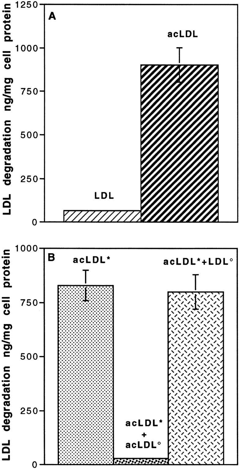
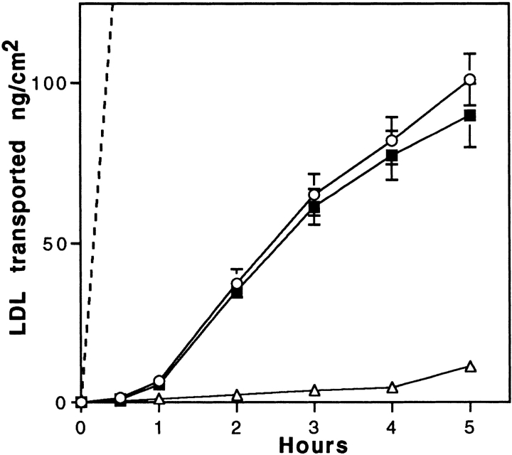
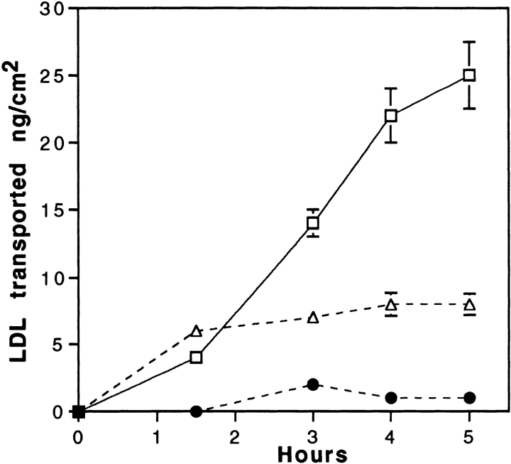
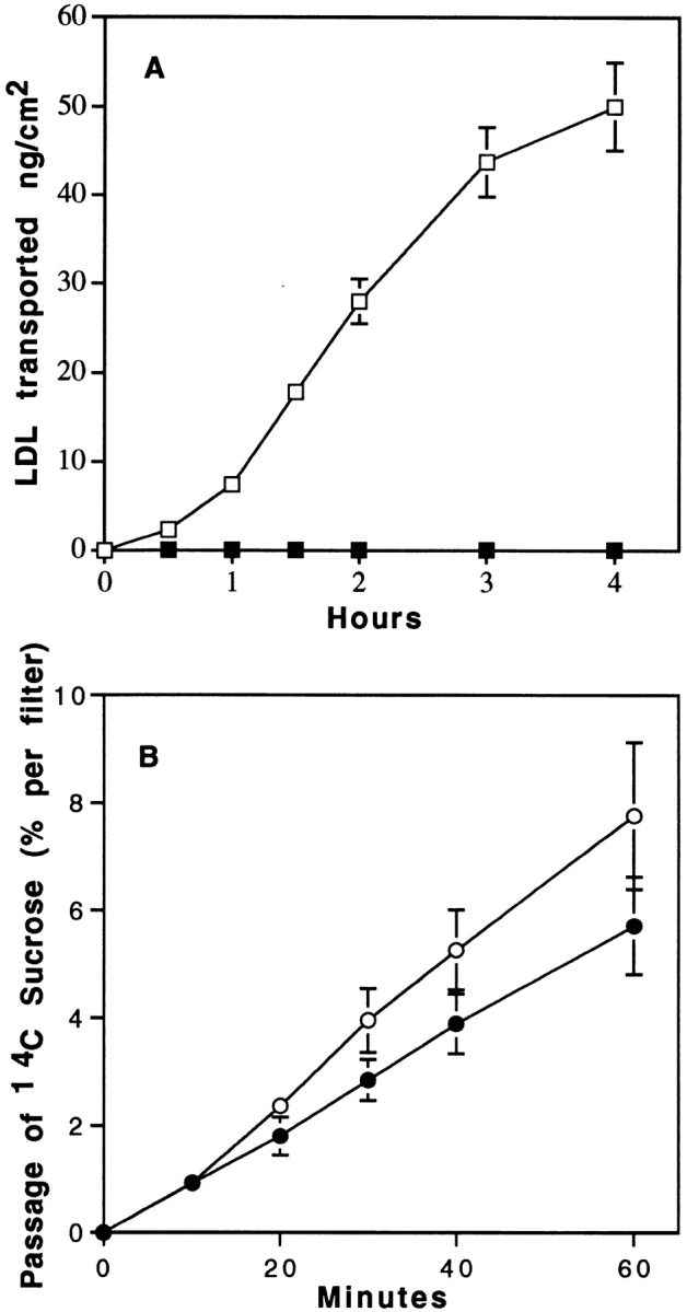
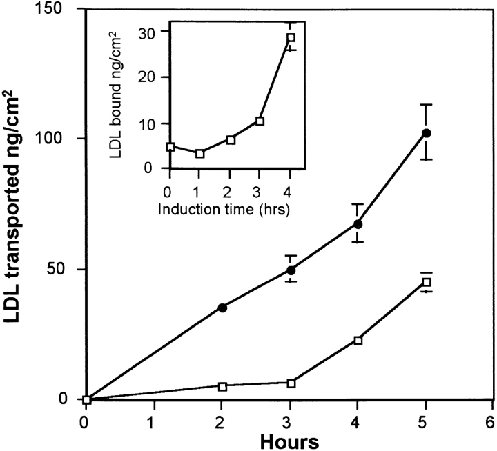
Lipoprotein transport across the blood-brain barrier (BBB) is of critical importance for the delivery of essential lipids to the brain cells. The occurrence of a low density lipoprotein (LDL) receptor on the BBB has recently been demonstrated. To examine further the function of this receptor, we have shown using an in vitro model of the BBB, that in contrast to acetylated LDL, which does not cross the BBB, LDL is specifically transcytosed across the monolayer. The C7 monoclonal antibody, known to interact with the LDL receptor-binding domain, totally blocked the transcytosis of LDL, suggesting that the transcytosis is mediated by the receptor. Furthermore, we have shown that cholesterol-depleted astrocytes upregulate the expression of the LDL receptor at the BBB. Under these conditions, we observed that the LDL transcytosis parallels the increase in the LDL receptor, indicating once more that the LDL is transcytosed by a receptor-mediated mechanism. The nondegradation of the LDL during the transcytosis indicates that the transcytotic pathway in brain capillary endothelial cells is different from the LDL receptor classical pathway. The switch between a recycling receptor to a transcytotic receptor cannot be explained by a modification of the internalization signals of the cytoplasmic domain of the receptor, since we have shown that LDL receptor messengers in growing brain capillary ECs (recycling LDL receptor) or differentiated cells (transcytotic receptor) are 100% identical, but we cannot exclude posttranslational modifications of the cytoplasmic domain, as demonstrated for the polymeric immunoglobulin receptor. Preliminary studies suggest that caveolae are likely to be involved in the potential transport of LDL from the blood to the brain.


Figures

Figure 1
Figure 1
Characterization of brain capillary EC monolayer grown on the upper face of a collagen-coated filter. (A) Phase contrast micrograph of confluent brain capillary ECs. (B) Endothelial staining with mitochondrion-selective dye, Mitotracker CMX Ros. (C) Staining with F-actin probe, bodipy–phallacidin (Molecular Probes, Inc). (D) Localization of tight junction-associated protein ZO-1 to the plasma membrane and nuclear staining by propidium iodine. Bar, 50 μm.
Figure 2
Figure 2
Electron and fluorescence microscopy of ECs incubated with Au16nm and DiI-labeled LDL. After a 5-min incubation with gold LDL at 37°C, the tracer marks the luminal plasma membrane (A). After a 45-min incubation, the gold-labeled probe is mainly detected in multivesicular bodies (C, arrow). Arrowheads indicate caveolar structure. (B) After 45 min of incubation, the fluorescent DiI-labeled LDL is endocytosed and found as small vesicles throughout the cells. Bars: (A and C) 0.3 μm; (B) 30 μm.
Figure 3
Figure 3
Degradation of LDL and acetylated LDL (acLDL) by brain capillary ECs. (A) Specific degradation of 125I-LDL and 125I-acLDL. Degradation was performed at the concentration of 50 μg/ml for 5 h at 37°C in the absence or the presence of a 20-fold excess of related, unlabeled lipoproteins. (B) Competition studies between labeled acLDL (*acLDL, 50 μg/ml; ░⃞ ) with unlabeled acLDL (acLDL°, 1 mg/ml; ░⃞ ) and unlabeled LDL (LDL°, 1 mg/ml; ▩ ). Determination of the degradation was carried out as described in Materials and Methods. Each point is a mean of three different filters, and the curves are representative of three series of experiments.
Figure 4
Figure 4
Transport of LDL across brain capillary EC monolayers. Passage of 125I-LDL (ng/cm2) through collagen-coated filters with (solid lines) or without (dashed line) brain capillary EC monolayer was carried out at 37°C. LDL was added to the upper side of the filter (50 μg/ml 125I-LDL, 1 mg/ml unlabeled LDL). Intact 125I-LDL transport from upper to lower sides of the filter was assessed by counting in a γ counter the acid-precipitable fractions of the lower compartments. Specific transport (▪) was calculated by subtracting the radioactivity obtained in the presence of native LDL (▵) from that obtained in the absence of native LDL (○). The data are expressed as ng of 125I-LDL transported per cm2, which refers to the surface area of the cells. Each point is a mean of three different filters, and the curves are representative of seven series of independent experiments.
Figure 5
Figure 5
Inhibition of LDL transport through EC monolayers by the C7 monoclonal antibody. Specific 125I-LDL transport experiment (35 μg/ml 125I-LDL, 700 mg/ml unlabeled LDL) was performed as described in Fig. 4, legend, either in presence of C7-IgG, 1 mg/ml (•), 0.1 mg/ml (▵), or AI-IgG, 1 mg/ml (□). Each point is a mean of three different filters, and the curves are representative of three series of independent experiments.
Figure 6
Figure 6
Effect of temperature on the transport of 125I-LDL (A) and sucrose (B) from apical to basal surfaces. Specific transport of LDL was performed as described in Fig. 4, legend. Transport of [14C]sucrose was expressed as the percentage of radioactivity crossing the brain capillary EC monolayer from the apical to the abluminal surfaces. 125I-LDL and sucrose transport were performed both at 37° (□, ○) and 4°C (▪, •). The data represent the means of three filters, and the curves are representative of two series of independent experiments.
Figure 7
Figure 7
LDL transport after induction of LDL receptor expression on brain capillary ECs by cholesterol-depleted astrocytes. Upregulation of LDL receptor on brain capillary ECs was performed in four phases as described in Materials and Methods. On the day of the experiment, brain capillary ECs in DME supplemented with 15% CS were cocultured for different times (1–4 h) at 37°C with cholesterol-depleted astrocytes in their 36-h incubating medium (induction phase). The increase of LDL receptor expression on ECs was studied by 125I-LDL binding at 4°C (inset). 125I-LDL transport studies were performed at 37°C, after 4 h induction and >5 h, as described in Fig. 4, legend. The induction phase was performed in the presence of either cholesterol- depleted (•) or noncholesterol-depleted (□) astrocytes. The data represent the means of three filters, and the curves are representative of three series of independent experiments.
Figure 8
Figure 8
DiI-LDL endocytosis after induction of LDL receptor expression on brain capillary ECs by cholesterol-depleted astrocytes. (A) Brain capillary ECs were incubated at 37°C for 45 min with DiI-LDL. After washing, the cells were fixed and processed for fluorescent microscopy as described in Materials and Methods. (B) The same experiment was carried out with “upregulated LDL receptor” ECs as described in Fig. 7, legend. Bar, 50 μm.
Figure 9
Figure 9
Effect of filipin on the endocytosis of LDL by brain capillary ECs. (A) The cells were pretreated with filipin (3 μg/ml) before the addition of DiI-LDL, and the endocytosis was performed as described in the Fig. 8, legend. (B) Immediately after filipin treatment, the cells were incubated in DME containing 20% CS for 30 min to reverse the effects of filipin before examining DiI-LDL endocytosis. (C and D) Endocytosis was carried out with brain capillary ECs in growing phase without (C) or with (D) pretreatment with filipin. Bar, 50 μm.

References

    1. Albers JJ, Tollefson JH, Wolfbauer G, Albright RE. Cholesteryl ester transfer protein in human brain. Int J Clin Lab Res. 1992;21:264–266. - PubMed
    1. Anderson RGW. Plasmalemmal caveolae and GPI-anchored membrane proteins. Curr Opin Cell Biol. 1993;5:647–652. - PubMed
    1. Aroeti B, Mostow KE. Polarized sorting of the polymeric immunoglobulin receptor in the exocytotic and endocytotic pathways is controlled by the same amino acids. EMBO (Eur Mol Biol Organ) J. 1994;13:2297–2304. - PMC - PubMed
    1. Basu SK, Goldstein J M, Anderson RGM, Brown MS. Degradation of cationized low density lipoprotein and regulation of cholesterol metabolism in homozygous familial hypercholesterolemia fibroblasts. Proc Natl Acad Sci USA. 1976;73:493–502. - PMC - PubMed
    1. Beisiegel U, Schneider WJ, Goldstein JL, Anderson RGW, Brown MS. Monoclonal antibodies to the low-density lipoprotein receptor as a probe to study of receptor mediated endocytosis and the genetics of familial hypercholesterolemia. J Biol Chem. 1981;256:11923–11931. - PubMed

Publication types