. 1997 Aug 25;138(4):891-9.
doi: 10.1083/jcb.138.4.891.

COOH terminus of occludin is required for tight junction barrier function in early Xenopus embryos

Affiliations

COOH terminus of occludin is required for tight junction barrier function in early Xenopus embryos

Y Chen et al. J Cell Biol. .

Abstract

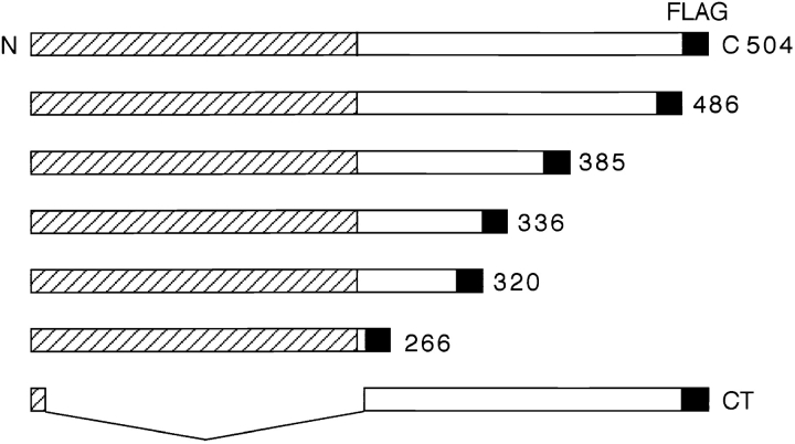
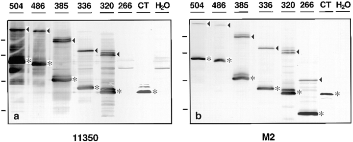
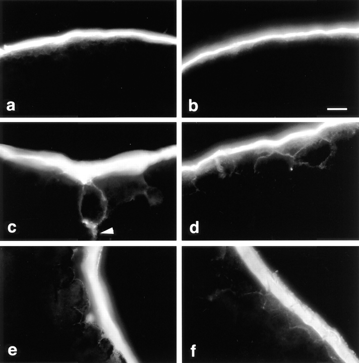
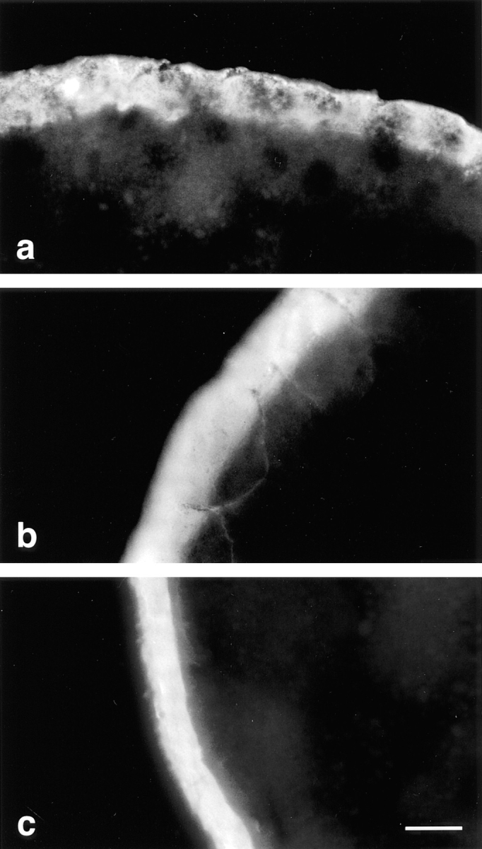
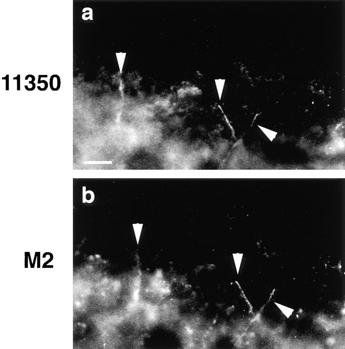
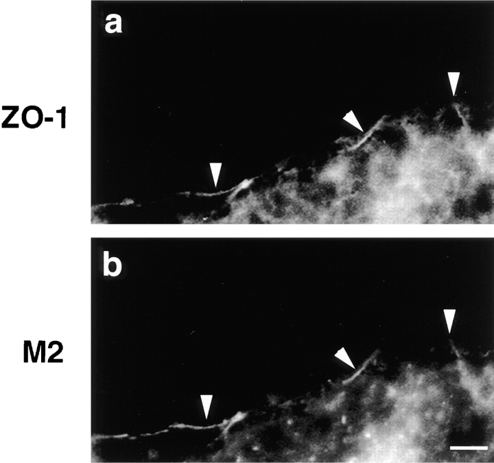
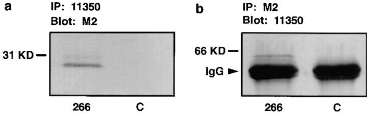
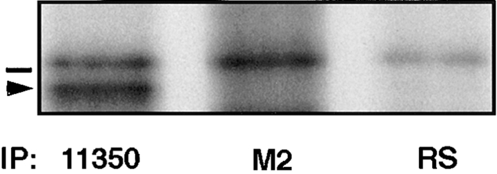
Occludin is the only known integral membrane protein localized at the points of membrane- membrane interaction of the tight junction. We have used the Xenopus embryo as an assay system to examine: (a) whether the expression of mutant occludin in embryos will disrupt the barrier function of tight junctions, and (b) whether there are signals within the occludin structure that are required for targeting to the sites of junctional interaction. mRNAs transcribed from a series of COOH-terminally truncated occludin mutants were microinjected into the antero-dorsal blastomere of eight-cell embryos. 8 h after injection, the full-length and the five COOH-terminally truncated proteins were all detected at tight junctions as defined by colocalization with both endogenous occludin and zonula occludens-1 demonstrating that exogenous occludin correctly targeted to the tight junction. Importantly, our data show that tight junctions containing four of the COOH-terminally truncated occludin proteins were leaky; the intercellular spaces between the apical cells were penetrated by sulfosuccinimidyl-6-(biotinamido) Hexanoate (NHS-LC-biotin). In contrast, embryos injected with mRNAs coding for the full-length, the least truncated, or the soluble COOH terminus remained impermeable to the NHS-LC-biotin tracer. The leakage induced by the mutant occludins could be rescued by coinjection with full-length occludin mRNA. Immunoprecipitation analysis of detergent-solubilized embryo membranes revealed that the exogenous occludin was bound to endogenous Xenopus occludin in vivo, indicating that occludin oligomerized during tight junction assembly. Our data demonstrate that the COOH terminus of occludin is required for the correct assembly of tight junction barrier function. We also provide evidence for the first time that occludin forms oligomers during the normal process of tight junction assembly. Our data suggest that mutant occludins target to the tight junction by virtue of their ability to oligomerize with full-length endogenous molecules.


Figures

Figure 1
Figure 1
Schematic drawing of full-length (504) and six truncated chick occludin proteins that were subcloned into expression vector SP64T. Each molecule has a FLAG epitope tag (black box) at the end of COOH terminus to distinguish it from Xenopus occludin. The shaded half of the molecule is predicted to traverse the membrane four times, and contains an NH2 terminus facing the cytoplasm, two extracellular loop domains, and one cytoplasmic loop domain. The unshaded half of the protein is predicted to face the cytoplasm. The numbers on the right indicate the total number of amino acids in each protein. CT, soluble NH2-terminal 20 amino acids fused to the predicted cytoplasmic tail; N, NH2 terminus; C, COOH terminus.
Figure 2
Figure 2
Western blot analysis of Xenopus oocytes injected with mRNAs corresponding to the occludin proteins diagrammed in Fig. 1. mRNAs were microinjected in the vegetal hemisphere of stage VI oocytes, and the injected oocytes were incubated at 17°C overnight. Oocyte homogenates were separated on 12% SDS-PAGE, transferred to Immobilon membranes, and then immunoblotted with either anti-occludin antibody 11350 (a) or with anti-FLAG monoclonal antibody M2 (b). The number on the top of each lane identifies each protein as named in Fig. 1. The asterisks in a and b indicate the predicted position of each expressed protein. With the exception of the soluble CT, the expressed proteins formed dimers as indicated by the arrowheads. Note that protein 266, with almost the entire COOH terminus deleted, was not recognized by 11350 antibody. H2O, control oocytes injected with water. Molecular weight markers (from top to bottom): 97.4, 66, 45, and 31 kD.
Figure 3
Figure 3
Functional assay of tight junctions by surface biotinylation in Xenopus embryos injected with chicken full-length or truncated occludins. 6 h after mRNA injection (2,000 cell blastula), the embryos were labeled by incubation in 1 mg/ml NHS-LC-biotin for 12 min at 10°C, washed, and then fixed in 3% formaldehyde in 80 mM sodium cacodylate. Frozen sections were stained with RITC- avidin and observed by fluorescence microscopy. In all embryos, NHS-LC-biotin reacts with molecules in the vitelline envelope, the subvitelline space, and the apical plasma membranes of the blastomeres, which together appear as a thick, continuous line at the surface of the embryo. The tight junctions in the embryos injected with 504 (a, the full-length) or 486 (b, the least COOH-terminally truncated) occludin mRNAs deny the biotin access to the intercellular spaces, and the basolateral membranes of the blastomeres are not stained. In the embryos injected with 385 (c), 336 (d) 320 (e), and 266 (f) (four COOH-terminally truncated occludin mRNAs), the biotin molecules penetrated into intercellular spaces demonstrating the disruption of the tight junction seal. Bar, 10 μm.
Figure 4
Figure 4
Coinjection of the full-length (504) occludin mRNA with the most COOH-terminally truncated occludin mRNA (266) rescued the tight junction leakage caused by injection of construct 266 alone. The experimental procedure was same as in Fig. 3. The frozen sections from the embryos injected with ∼2 pg of 504 (a), 266 (b), or 504 and 266 (c) construct mRNA were stained with RITC-avidin. The extraembryonic space is toward the top of figure in a, and left in b and c. No leak was seen in the embryo infected with 504 mRNA (a). Biotin molecules clearly penetrated through the tight junctions and labeled the intercellular spaces between the apical cells of embryo injected with 266 mRNA (b). However, there was no detectable leak in the embryo injected with both 504 and 266 mRNA (c). Bar, 15 μm.
Figure 5
Figure 5
Double labeling of frozen sections of Xenopus embryos with anti-occludin 11350 (a) and anti-FLAG M2 (b). 4 nl (∼10 pg) of mutant 266 mRNA were microinjected into the anterior, dorsal blastomere of eight-cell stage embryo and incubated for 8 h at room temperature before freezing. The arrowheads indicate the colocalization of Xenopus full-length occludin and chicken truncated occludin at the junctional complexes. Bar, 10 μm.
Figure 6
Figure 6
Immunocolocalization of Xenopus ZO-1 with the protein directed by mutant 266, the most truncated occludin in Xenopus embryos. Frozen sections were made of embryos injected with mutant 266 mRNA and stained with either anti–ZO-1 (a) or with anti-FLAG M2 (b). The arrowheads indicate the colocalization of both proteins at the junctional complexes. Bar, 10 μm.
Figure 7
Figure 7
The interaction of Xenopus occludin with chicken occludin in vivo assayed by immunoprecipitation. The embryos were microinjected with the most COOH-terminally truncated occludin mRNA (266; a and b, left lanes) or water (C; a and b, right lanes) and incubated at room temperature for 10 h. After homogenization, the samples were centrifuged at 100,000 g for 30 min at 4°C. The membrane pellet was solubilized in modified RIPA buffer (see Materials and Methods) and centrifuged at 100,000 g for 1 h at 4°C. The resulting supernatant was immunoprecipitated with anti-occludin 11350 (a) or anti-FLAG M2 (b) overnight at 4°C. Immunoprecipitates were immunoblotted with anti-FLAG M2 (a) or anti-occludin 11350 (b). A specific band corresponding to the chicken truncated occludin (compare with Fig. 2 b, lane 266) was present in a (left lane) after immunoprecipitated with anti-occludin 11350. The Xenopus full-length occludin was observed at b (left lane) after immunoprecipitated with anti–FLAG M2. The two thick bands in b were IgG heavy chain.
Figure 8
Figure 8
Coimmunoprecipitation of the mutant with endogenous occludin was not the result of exchange of monomers between detergent-solubilized oligomeric assemblies. Confluent A6 cell monolayers were metabolically labeled with 150 μCi/ml [35S]methionine/cysteine for 20 h at 27°C, solubilized under nondenaturing conditions, and then mixed with similarly detergent-solubilized supernatant from Xenopus embryos injected with 266 construct mRNA at the two-cell stage and incubated for 10 h at room temperature. After immunoprecipitation with anti-occludin 11350 (left lane), anti-FLAG M2 (middle lane), or preimmune rabbit serum (right lane), samples were separated on 12% SDS-PAGE and autoradiographed. 35S-labeled occludin could not be immunoprecipitated with anti-FLAG M2 (middle lane), demonstrating that mixing of the mutant with wild-type molecules after extraction did not result in detergent-mediated exchange of protein monomers. The bar above the arrowhead indicates the molecular weight marker 66 kD.

References

    1. Ando-Akatsuka Y, Saitou M, Hirase T, Kishi M, Sakakibara A, Itoh M, Yonemura S, Furuse M, Tsukita S. Interspecies diversity of the occludin sequence: cDNA cloning of human, mouse, dog, and rat-kangaroo homologues. J Cell Biol. 1996;133:43–47. - PMC - PubMed
    1. Balda MS, Whitney JA, Flores C, Gonzalez S, Cereijido M, Matter K. Functional dissociation of paracellular permeability and transepithelial electrical resistance and disruption of the apical-basolateral intramembrane diffusion barrier by expression of a mutant tight junction membrane protein. J Cell Biol. 1996;134:1031–1049. - PMC - PubMed
    1. Cereijido M, Ponce A, Gonzalez-Mariscal L. Tight junctions and apical/basolateral polarity. J Membr Biol. 1989;110:1–9.
    1. Citi S, Sabanay H, Jakes R, Geiger B, Kendrick-Jones J. Cingulin, a new peripheral component of tight junctions. Nature (Lond) 1988;333:272–276. - PubMed
    1. Claude P, Goodenough DA. Fracture faces of zonulae occludentes from “tight” and “leaky” epithelia. J Cell Biol. 1973;58:390–400. - PMC - PubMed

Publication types

MeSH terms