. 1997 Aug 1;11(15):1925-37.
doi: 10.1101/gad.11.15.1925.

Null mutation of mCOUP-TFI results in defects in morphogenesis of the glossopharyngeal ganglion, axonal projection, and arborization

Affiliations

Null mutation of mCOUP-TFI results in defects in morphogenesis of the glossopharyngeal ganglion, axonal projection, and arborization

Y Qiu et al. Genes Dev. .

Abstract

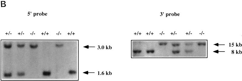
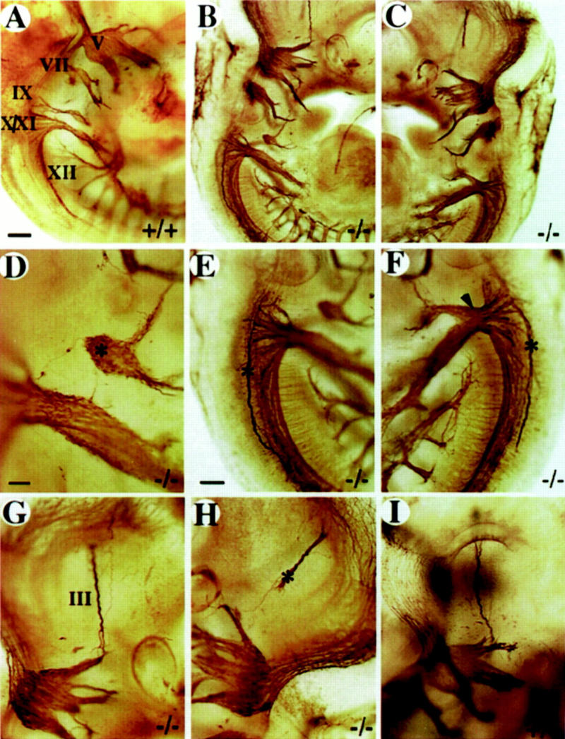
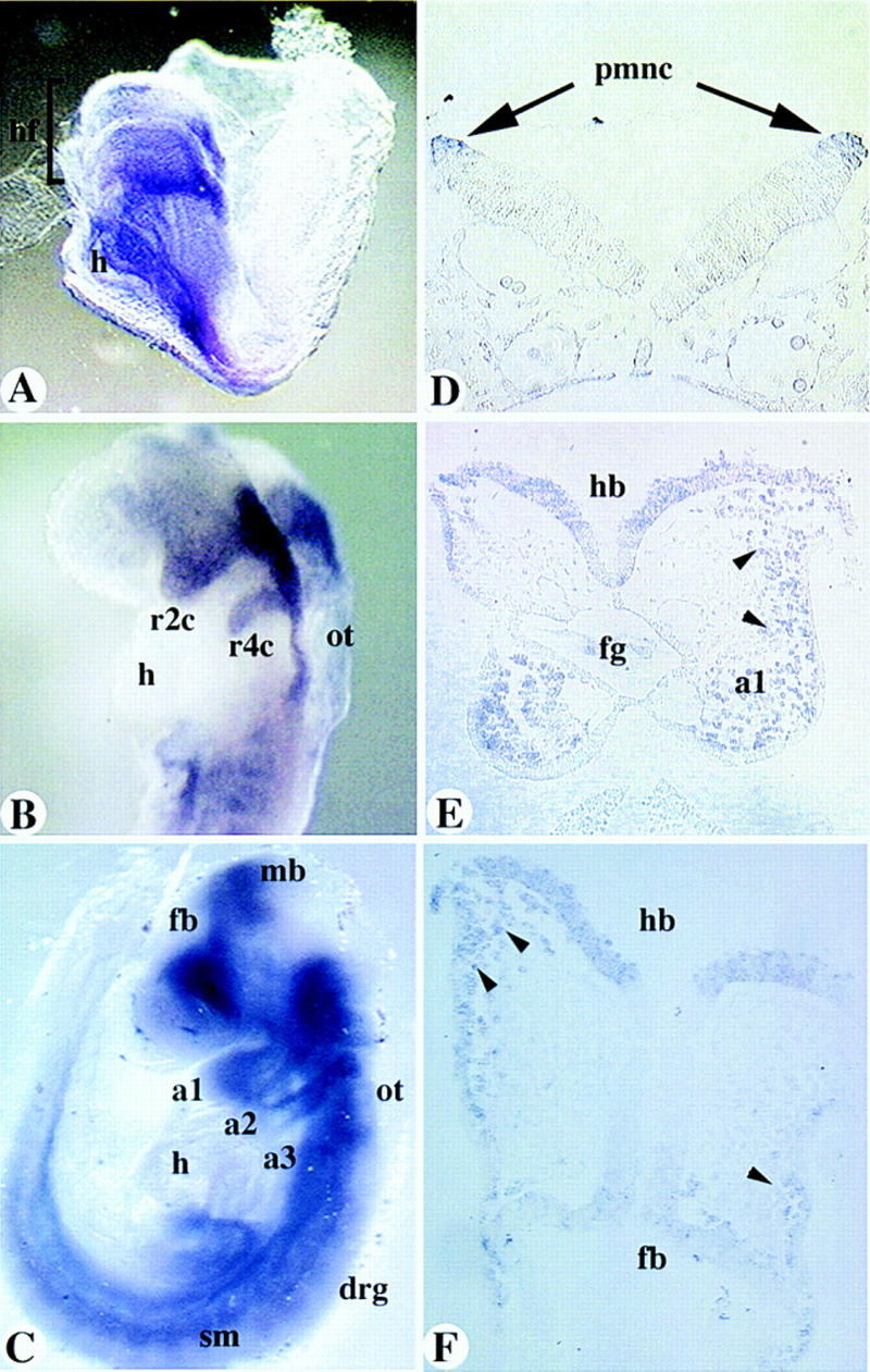
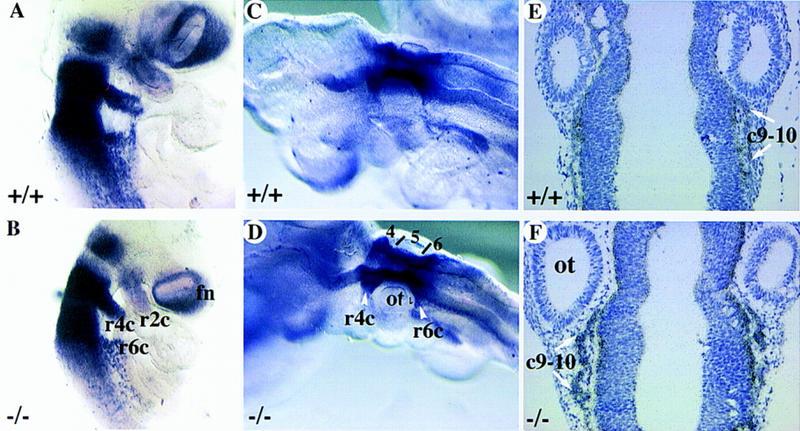
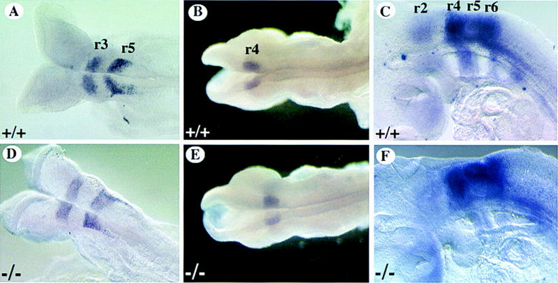
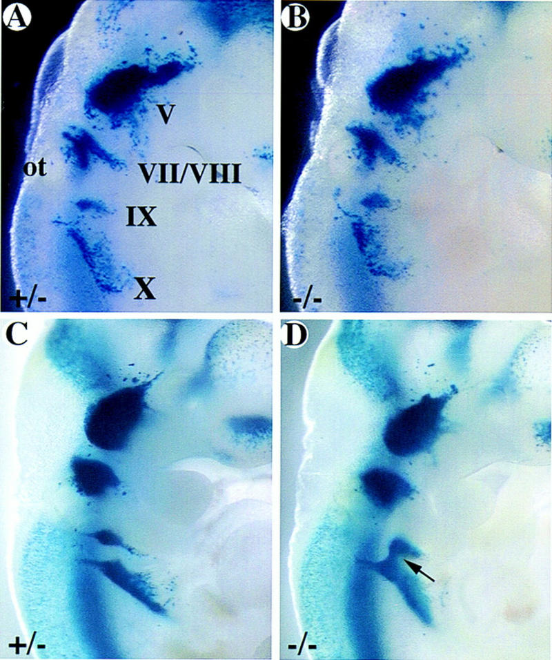
The COUP-TFs are orphan members of the steroid/thyroid hormone receptor superfamily. Multiple COUP-TF members have been cloned and they share a high degree of sequence homology between species as divergent as Drosophila and humans, suggesting a conservation of function through evolution. The COUP-TFs are highly expressed in the developing nervous systems of several species examined, indicating their possible involvement in neuronal development and differentiation. In the mouse, there are two very homologous COUP-TF genes (I and II) and their expression patterns overlap extensively. To study the physiological function of mCOUP-TFI, a gene-targeting approach was undertaken. We report here that mCOUP-TFI null animals die perinataly. Mutant embryos display an altered morphogenesis of the ninth cranial ganglion and nerve. The aberrant formation of the ninth ganglion is most possibly attributable to extra cell death in the neuronal precursor cell population. In addition, at midgestation, aberrant nerve projection and arborization were oberved in several other regions of mutant embryos. These results indicate that mCOUP-TFI is required for proper fetal development and is essential for postnatal development. Furthermore, mCOUP-TFI possesses vital physiological functions that are distinct from mCOUP-TFII despite of their high degree of homology and extensive overlapping expression patterns.


Figures

Figure 1
Figure 1
Targeted disruption of the mCOUP-TFI locus. (A) Targeting strategy. The mCOUP-TFI locus is shown on top with exons I–III in boxes. The solid region represents open reading frame and open boxes represents the 5′- and 3′-untranslated regions. The hatched regions represent probes used in Southern blot analysis. The replacement vector includes a PGK-neo gene and a tk gene. The directions of transcription are indicated by arrows. The shaded box represents pBluescript vector. The left and right arms are indicated by dashed lines. The SacI site at the right end of the left arm was destroyed during subcloning. The correctly recombined locus is shown at the bottom. Restriction sites are indicated above the lines. (P) PstI; (S) SmaI; (X) XbaI; (R1) EcoRI; (B) BamHI; (SL) SalI. (B) Southern blot analysis on a representative litter from a mCOUP-TFI heterozygote intercross. Genomic DNA was digested with SacI and hybridized with the 5′ probe. A 1.6-kb and a 3.0-kb band were generated from wild type and mutant alleles, respectively. For the 3′ probe, BamHI was used. The 8-kb and the 15-kb fragments were generated from the wild type and mutant allele, respectively. (C) Northern analysis of RNA from E13.5 wildtype (+/+), heterozygote (+/−), and homozygote embryos (−/−). A 700-bp PstI–ScaI fragment mainly containing the 3′ untranslated region of mCOUP-TFI was used as the probe. Note the absence of mCOUP-TFI mRNA in the homozygote. GAPDH was used as an internal control and is shown at bottom.
Figure 1
Figure 1
Targeted disruption of the mCOUP-TFI locus. (A) Targeting strategy. The mCOUP-TFI locus is shown on top with exons I–III in boxes. The solid region represents open reading frame and open boxes represents the 5′- and 3′-untranslated regions. The hatched regions represent probes used in Southern blot analysis. The replacement vector includes a PGK-neo gene and a tk gene. The directions of transcription are indicated by arrows. The shaded box represents pBluescript vector. The left and right arms are indicated by dashed lines. The SacI site at the right end of the left arm was destroyed during subcloning. The correctly recombined locus is shown at the bottom. Restriction sites are indicated above the lines. (P) PstI; (S) SmaI; (X) XbaI; (R1) EcoRI; (B) BamHI; (SL) SalI. (B) Southern blot analysis on a representative litter from a mCOUP-TFI heterozygote intercross. Genomic DNA was digested with SacI and hybridized with the 5′ probe. A 1.6-kb and a 3.0-kb band were generated from wild type and mutant alleles, respectively. For the 3′ probe, BamHI was used. The 8-kb and the 15-kb fragments were generated from the wild type and mutant allele, respectively. (C) Northern analysis of RNA from E13.5 wildtype (+/+), heterozygote (+/−), and homozygote embryos (−/−). A 700-bp PstI–ScaI fragment mainly containing the 3′ untranslated region of mCOUP-TFI was used as the probe. Note the absence of mCOUP-TFI mRNA in the homozygote. GAPDH was used as an internal control and is shown at bottom.
Figure 2
Figure 2
Whole-mount immunohistochemistry on wild-type and homozygote embryos using 2H3 anti-neurofilament antibody showing the progression of cranial ganglion formation. (A,C) Wild-type and (B,D) mutant embryos at E9.5 and E10.5, respectively. Dorsal to the left, ventral to the right. Note the ganglion IX in two different stages of embryos. Arrows in A and C point to the nerve projections between the ninth ganglion and the hindbrain. Arrows in B and D point to the absence of nerve projection between the ninth ganglion and the hindbrain. The arrowheads in B and D point to the connections between ganglia IX and X. (V) Trigeminal ganglion; (VII) facial ganglion; (IX) glossopharyngeal ganglion; (X) vagus ganglion; (XI) accessory ganglion; (XII) hypoglossal nerve. ot, otic vesicle. Bar, 100 μm.
Figure 8
Figure 8
Whole-mount analysis of axonal projections. Multiple defects in cranial nerve projections are detected in severely affected embryos. (A) wild-type E10.5 embryo. The right (B) and the left (C) side of a E10.5 mutant embryo. (D–F) Enlargement of cranial nerve IX–XII region from B and C. Note the isolated ganglionic mass (asterisk in D) at the position of the ninth ganglion, the abnormal projection of a single nerve fiber toward the hindbrain (D) and the abnormal axonal bundles (asterisk in E,F) on both sides of the hindbrain. Note the extensive fusion of nerve IX and X on the left side of the animal (arrowhead in F). (G,H) Enlargement of the oculomotor nerve (III) region in B and C. Note the abnormal projection of nerve III on the left side (asterisk in H). (III) Oculomotor nerve; (V) trigeminal ganglion; (VII) facial ganglion; (IX) glossopharyngeal ganglion; (X) vagus ganglion; (XI) accessory ganglion; (XII) hypoglossal nerve. (A–C) Bar, 100 μm; (E–H) bar, 50 μm; (D) bar, 20 μm.
Figure 3
Figure 3
Expression of mCOUP-TFI. (A–C) Whole-mount in situ hybridization of mCOUP-TFI in E8.0 (A), E8.5 (B), and 9.0 (C) embryos, respectively. (D–F) Sections of a whole-mount stained E8.5 embryo. (a1, a2, a3) branchial arch 1, 2, and 3, respectively; (drg) dorsal root ganglia; (fb) forebrain; (h) heart; (hb) hindbrain; (hf) head fold; (fg) foregut; (mb) midbrain; (ot) otic vesicle; (pmnc) premigratory neural crest cells; (r2c, r4c) neural crest cells from r2, r4, respectively; (sm) somite. Arrows in D point to premigratory neural crest cells (NCC). Arrowheads in E and F indicate migrating NCC that are mCOUP-TFI positive.
Figure 5
Figure 5
Analysis of cranial NCC migration in mCOUP-TFI mutants. Wild-type (A–C) or mutant embryos (D–F) were hybridized with CRABP I antisense probe to examine the migration of the neural crest cells. Whole-mount in situ hybridization on E9.0 (A,D) and E9.5 (B,E) embryos. (C,F) in situ hybridization with CRABP I antisense probe on sections of late E9.5 embryos. (c9–c10) Neural crest cells for ganglia IX and X; (fn) frontal nasal mesenchyme; (r) rhombomere; (r2c, r4c, r6c) neural crest cells from r2, r4, r6, respectively; (ot) otic vesicle; (4, 5, and 6) r4, r5, and r6, respectively.
Figure 4
Figure 4
Expression of rhombomere-specific genes in mCOUP-TFI mutants. Whole-mount in situ hybridization was performed on wild-type (A–C) or mutant embryos (D–F) with rhombomere specific markers. Note expression is unchanged for mutants. (A,D) Krox-20 expression in r3 and r5 of E8.5 embryos. (B,E) Hoxb-1 expression in r4 of E9.0 embryos. (C,F) CRABP I expression strong in r4–r6, weak in r2 of E9.5 embryos.
Figure 6
Figure 6
Developmental progression of the formation of ganglion IX as visualized by X-gal staining. mCOUP-TFI wild-type (A,C) and mutant (B,D) embryos at E9.5 (A,B) and E10.5 (C,D) were stained with X-gal. A and B were among the embryos used for apoptosis study; C and D were among the embryos used for neuronal counting, as shown in Table 2. (V) Trigeminal ganglion; (VII/VIII) facial and acoustic ganglia; (IX) glossopharyngeal ganglion; (X) vagus ganglion. Arrow indicates the fusion between ganglia IX and X in the mutant embryo.
Figure 7
Figure 7
Excessive cell death in mCOUP-TFI mutant embryos. mCOUP-TFI mutant (A–D) and wild-type (E–H) embryos at E9.5 were stained with X-gal, sectioned, and TUNEL assay was performed. Every other section in the region of interest is presented. Sections at top are more dorsal than the sections at the bottom. Note that the left side of the mutant embryo shows much more pronounced cell death than in the wild type. (ot) Otic vesicle; (VII/VIII) facial and acoustic ganglia; (X) vagus ganglion. Note that IX marks a position dorsal to ganglion IX proper, which lies further ventral. (Arrows) Apoptotic cells.
Figure 9
Figure 9
mCOUP-TFI mutant embryos display reduced arborization of axonal trees. Whole-mount immunohistochemistry of wild-type (A,C) and mutant E11.5 fetuses (B,D) using 2H3 anti-neurofilament antibody. (A,B) Sagittal views at the level of posterior hindbrain and anterior somite region showing arborization of the first several spinal nerves. Note the reduced arborization in the mutant fetuses. (C,D) Higher magnification of the ophthalmic branch of the trigeminal nerve. Arrowheads point to the corresponding branching points in the wild-type and mutant embryos. Note the extensive branchings in the wild type. Also, note the less extent of arborization in the bracketed region.

References

    1. Behringer RR, Crotty DA, Tennyson VM, Brinster RL, Palmiter RD, Wolgemuth DJ. Sequences 5′ of the homeobox of the Hox-1.4 gene direct tissue-specific expression of LacZ during mouse development. Development. 1993;117:823–833. - PubMed
    1. Chazaud CM, Oulad-Abdelghani M, Bouillet P, Decimo D, Chambon P, Dolle P. AP-2.2, a novel gene related to AP-2, is expressed in the forebrain, limbs and face during mouse embryogenesis. Mech Dev. 1996;54:83–94. - PubMed
    1. Church GM, Gilbert W. Genomic sequencing. Proc Natl Acad Sci. 1984;81:1991–1995. - PMC - PubMed
    1. Cooney A, Tsai S, O’Malley BW, Tsai M-J. Chicken ovalbumin upstream promoter transcription factor (COUP-TF) dimers bind to different GGTCA response elements, allowing COUP-TF to repress hormonal induction of the vitamin D3, thyroid hormone, and retinoic acid receptors. Mol Cell Biol. 1992;12:4153–4163. - PMC - PubMed
    1. Cooney A, Leng X, Tsai S, O’Malley BW, Tsai M-J. Multiple mechanisms of chicken ovalbumin upstream promoter transcription factor-dependent repression of transactivation by the vitamin D, thyroid hormone, and retinoic acid receptors. J Biol Chem. 1993;268:4152–4160. - PubMed

Publication types

MeSH terms