. 1997 Aug 1;11(15):1987-98.
doi: 10.1101/gad.11.15.1987.

PPARgamma induces cell cycle withdrawal: inhibition of E2F/DP DNA-binding activity via down-regulation of PP2A

Affiliations

PPARgamma induces cell cycle withdrawal: inhibition of E2F/DP DNA-binding activity via down-regulation of PP2A

S Altiok et al. Genes Dev. .

Abstract

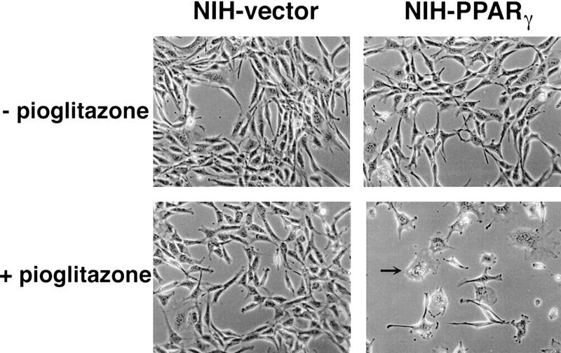
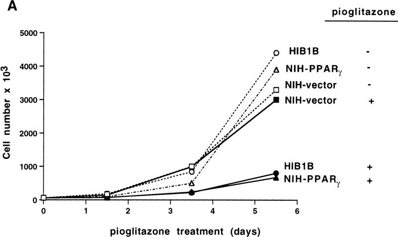
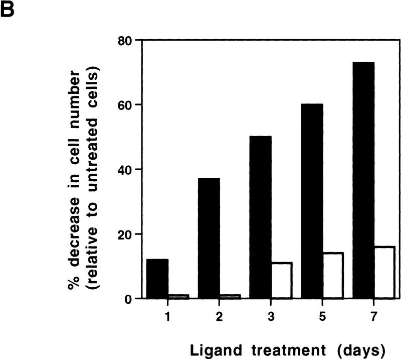
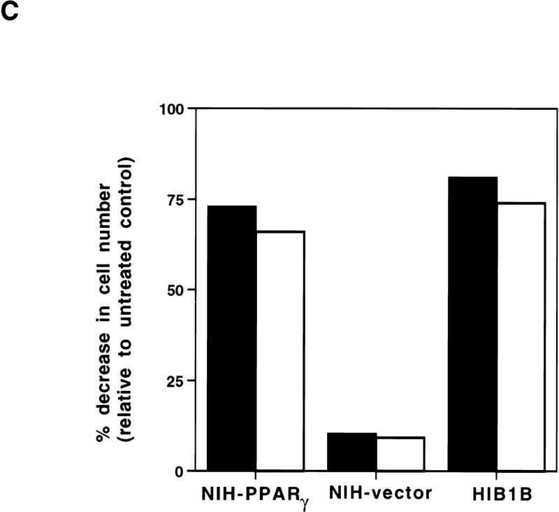
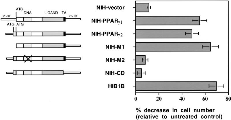
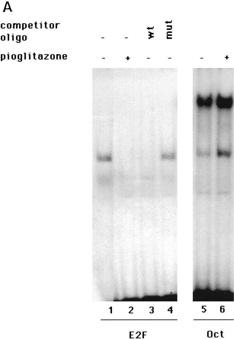
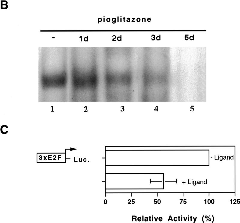
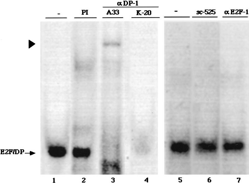
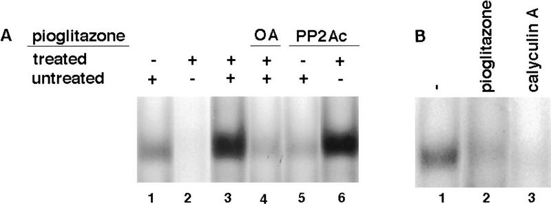
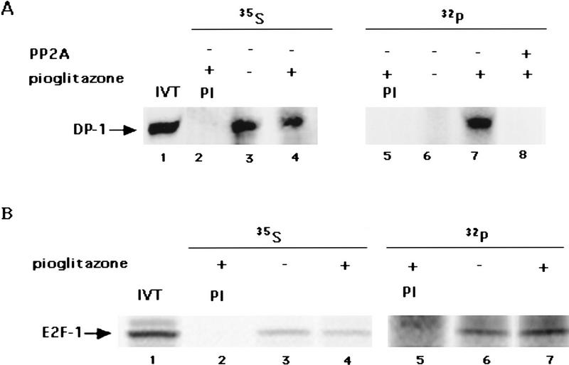
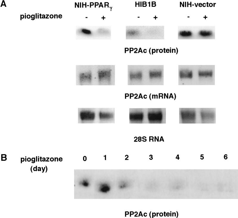
PPAR gamma is an adipose-selective nuclear hormone receptor that plays a key role in the control of adipocyte differentiation. Previous studies indicated that activation of ectopically expressed PPAR gamma induces differentiation when cells have ceased growth because of confluence. We show here that ligand activation of PPAR gamma is sufficient to induce growth arrest in fibroblasts and SV40 large T-antigen transformed, adipogenic HIB1B cells. Cell cycle withdrawal is accompanied by a decrease in the DNA-binding and transcriptional activity of the E2F/DP complex, which is attributable to an increase in the phosphorylation of these proteins, especially DP-1. This effect is a consequence of decreased expression of the catalytic subunit of the serine-threonine phosphatase PP2A. These data suggest an important role for PP2A in the control of E2F/DP activity and a new mode of cell cycle control in differentiation.


Figures

Figure 1
Figure 1
Pioglitazone stimulates growth arrest as well as adipose differentiation of NIH-3T3 cells expressing PPARγ ectopically. NIH-3T3 cells were infected with the retrovirus containing PPARγ expression vector (NIH–PPARγ) or with the empty vector (NIH-vector). After selection in puromycin, cells were pooled and cultured with or without pioglitazone (5 μm) for 5 days. Arrow shows a differentiated adipocyte containing lipid drops in the cytoplasm.
Figure 2
Figure 2
Growth of NIH–PPARγ, NIH-vector, or HIB1B cells in the presence or absence of PPARγ ligands. The same number of NIH–PPARγ, NIH-vector, or HIB1B cells were cultured either in the presence or absence of PPARγ ligands. Cell numbers were determined at the indicated time points. (A) Cumulative growth of cells untreated or treated with 5 μm pioglitazone is shown. (B) Percent decrease of the cell numbers in the pioglitazone-treated plates (solid bars) relative to the untreated plates (open bars) is shown. (C) Exponentially growing cells were treated without or with two members of thiozolidinediones, pioglitazone (5 μm) (solid bars) or BRL49653 (1 μm) (open bars) for 5 days and cell numbers were determined. The effect of ligands on cell growth is represented as percentage decrease in cell numbers in the treated plates relative to untreated control plates.
Figure 2
Figure 2
Growth of NIH–PPARγ, NIH-vector, or HIB1B cells in the presence or absence of PPARγ ligands. The same number of NIH–PPARγ, NIH-vector, or HIB1B cells were cultured either in the presence or absence of PPARγ ligands. Cell numbers were determined at the indicated time points. (A) Cumulative growth of cells untreated or treated with 5 μm pioglitazone is shown. (B) Percent decrease of the cell numbers in the pioglitazone-treated plates (solid bars) relative to the untreated plates (open bars) is shown. (C) Exponentially growing cells were treated without or with two members of thiozolidinediones, pioglitazone (5 μm) (solid bars) or BRL49653 (1 μm) (open bars) for 5 days and cell numbers were determined. The effect of ligands on cell growth is represented as percentage decrease in cell numbers in the treated plates relative to untreated control plates.
Figure 3
Figure 3
Transcription factor activity of PPARγ is required for its negative regulatory function on cell growth. (Left) Schematic representations of wild-type PPARγ1, PPARγ2, or mutant PPARγ2 cDNAs. NIH–M1 cells contain PPARγ expression vector, which lacks the first 127 amino acids located in the amino terminus. NIH–M2 cells express a PPARγ2 receptor in which cystein residues at the DNA-binding domain at positions 156 and 159 have been changed to serine. NIH–CD cells express a truncated form of PPARγ2, which lacks the conserved carboxyl-terminal transactivation domain. (Right) Effects of pioglitazone treatment on the growth rate of cells expressing wild-type or mutant forms of PPARγ. Cell numbers were determined after 5 days treatment without or with 5 μm pioglitazone. Decrease in the cell number in treated plates was represented as relative change to untreated control plates. The data represent the average of at least three independent experiments.
Figure 4
Figure 4
PPARγ-induced growth arrest is accompanied by a strong decrease in the E2F/DP DNA-binding activity. (A) HIB1B cells were treated as described in the legend to Fig. 2. Whole cell extracts were prepared and electrophoretic mobility-shift assays were performed by using E2F or Oct oligonucleotides as probe (see Materials and Methods). For competition experiments a 100-fold molar excess of unlabeled wild-type (wt) or mutant (mut) oligonucleotides were used. (B) HIB1B cells were treated for various times with 5 μm pioglitazone. Whole cell extracts were prepared and electrophoretic mobility-shift assays were performed with E2F probe. (C) HIB1B cells were transfected transiently with a luciferase reporter gene linked to three tandemly repeated E2F-binding sites. All transfections included a plasmid directing the expression of β-galactosidase under the control of β-actin promoter. Twenty-four hours after transfection, cells were cultured for an additional 48 hr in the presence or absence of 5 μm pioglitazone. Cells were harvested and assayed for luciferase activity, which was normalized for variable transfection efficiencies by correcting for β-galactosidase activity. Decrease in the luciferase activity in pioglitazone-treated cells is shown as a change relative to untreated control plates. Results are the average of six independent transfection experiments.
Figure 4
Figure 4
PPARγ-induced growth arrest is accompanied by a strong decrease in the E2F/DP DNA-binding activity. (A) HIB1B cells were treated as described in the legend to Fig. 2. Whole cell extracts were prepared and electrophoretic mobility-shift assays were performed by using E2F or Oct oligonucleotides as probe (see Materials and Methods). For competition experiments a 100-fold molar excess of unlabeled wild-type (wt) or mutant (mut) oligonucleotides were used. (B) HIB1B cells were treated for various times with 5 μm pioglitazone. Whole cell extracts were prepared and electrophoretic mobility-shift assays were performed with E2F probe. (C) HIB1B cells were transfected transiently with a luciferase reporter gene linked to three tandemly repeated E2F-binding sites. All transfections included a plasmid directing the expression of β-galactosidase under the control of β-actin promoter. Twenty-four hours after transfection, cells were cultured for an additional 48 hr in the presence or absence of 5 μm pioglitazone. Cells were harvested and assayed for luciferase activity, which was normalized for variable transfection efficiencies by correcting for β-galactosidase activity. Decrease in the luciferase activity in pioglitazone-treated cells is shown as a change relative to untreated control plates. Results are the average of six independent transfection experiments.
Figure 5
Figure 5
DP-1 is a major component of the endogenous E2F/DP DNA-binding activity. Extracts from exponentially growing HIB1B cells were incubated with preimmune serum (PI) or with the various antibodies as indicated here and then subjected to electrophoretic mobility-shift assays using a 32P-labeled E2F probe. The respective antibodies were as follows: K-20, an α-DP-1 polyclonal antibody (Santa Cruz sc-610x); A33, a rabbit polyclonal antiserum raised against DP-1; α-E2F-1, a mouse monoclonal antibody (Santa Cruz sc-251x); sc-525, an unrelated control antibody prepared against the GADD45 protein (Santa Cruz). Arrowhead shows the supershift.
Figure 6
Figure 6
PP2Ac is able to restore decreased E2F/DP DNA binding activity in pioglitazone-treated cells. (A) Whole cell extracts prepared from untreated (lane 1) or pioglitazone (5 μm)-treated (lane 2) HIB1B cells were used either directly or first mixed in the absence (lane 3) or presence (lane 4) of 3 nm okadaic acid (OA) and then used for gel mobility-shift assays to determine E2F/DP DNA-binding activity. Extracts from ligand untreated (lane 5) or treated (lane 6) HIB1B cells were treated with 0.5 unit of PP2Ac (GIBCO BRL) for 30 min at room temperature and electrophoretic mobility-shift assays were carried out by using E2F oligonucleotide as probe. The data shown represent the results of a single experiment. Exactly identical results were obtained in another independent experiment. (B) Calyculin A treatment leads to a decrease in the E2F/DP DNA-binding activity. HIB1B cells were treated with 5 μm pioglitazone or with 0.1 nm calyculin A for 3 days. Whole cell extracts were prepared from HIB1B cells treated as described above, and gel mobility-shift assays were performed to determine the DNA-binding activity of endogenous E2F/DP.
Figure 7
Figure 7
Activation of PPARγ induces phosphorylation of DP-1 but not E2F-1. HIB1B cells with or without pioglitazone treatment for 3 days were metabolically labeled with [32P]orthophosphate or with [35S]methionine. Whole cell lysates were prepared and then subjected to immunoprecipitation using preimmune serum (PI), antiserum specific for DP-1, or antibody prepared for HA. (A) DP-1 immunoprecipitates were boiled in 1% SDS, and 32P-labeled DP-1 was reimmunoprecipitated with DP-1 antibody. Immunoprecipitates prepared from PPARγ-activated cells were either tested directly (lane 4) or after treatment with PP2Ac for 20 min at room temperature (lane 5). Lane 1 shows 35S-labeled in vitro translated DP-1. (B) Lysates of metabolically labeled HIB1B cells ectopically expressing HA–E2F-1 were subjected to preimmuneserum (PI) or anti-HA antibody MAb 12CA5. 35S-Labeled, in vitro-translated HA–E2F-1 is shown in lane 1. Samples were analyzed by SDS–gel electrophoresis under reducing conditions. Mobilities of DP-1 and E2F-1 are indicated by arrows.
Figure 8
Figure 8
Activation of PPARγ leads to decreased PP2Ac but not mRNA. (A) Exponentially growing NIH–PPARγ, NIH-vector, or HIB1B cells were cultured in the absence or presence of 5 μm pioglitazone for 5 days. Total RNA or whole cell lysates were then prepared. For Northern analysis total RNA (10 μg) was blotted to nylon membrane and hybridized with 32P-labeled mouse PP2Ac prepared by RT-PCR from NIH-3T3 cells. An equivalent amount of intact RNA was run in each lane as indicated as 28S RNA. The lysates were equalized for protein content and processed for Western blotting with anti-PP2Ac antibody. (B) HIB16B cells were treated for various times with 5 μm pioglitazone. Lysates were prepared and processed for Western blotting.

References

    1. Beijersbergen RL, Kerkhoven RM, Zhu L, Carlee L, Mathijs FM, Bernards R. E2F-4, a new member of the E2F gene family, has oncogenic activity and associates with p107 in vivo. Genes & Dev. 1994;8:2680–2690. - PubMed
    1. Brun RP, Tontonoz P, Forman BM, Ellis R, Chen J, Evans RM, Spiegelman BM. Differential activation of adipogenesis by multiple PPAR isoforms. Genes & Dev. 1996;10:974–984. - PubMed
    1. Cao L, Faha B, Dembski M, Tsai LH, Harlow E, Dyson N. Independent binding of the retinoblastoma protein and p107 to the transcription factor E2F. Nature. 1992;355:176–179. - PubMed
    1. Cao Z, Umek RM, McKnight SL. Regulated expression of three C/EBP isoforms during adipose conversion of 3T3-L1 cells. Genes & Dev. 1991;5:1538–1552. - PubMed
    1. Chirgwin JM, Przybyla AE, MacDonald RJ, Rutter WJ. Isolation of biologically active ribonucleic acid from sources enriched in ribonuclease. Biochemistry. 1979;18:5294–5299. - PubMed

Publication types

MeSH terms

LinkOut - more resources