. 1997 Aug 29;186(5):655-63.
doi: 10.1084/jem.186.5.655.

Polymerase chain reaction selects a novel disintegrin proteinase from CD40-activated germinal center dendritic cells

Affiliations

Polymerase chain reaction selects a novel disintegrin proteinase from CD40-activated germinal center dendritic cells

C G Mueller et al. J Exp Med. .

Abstract

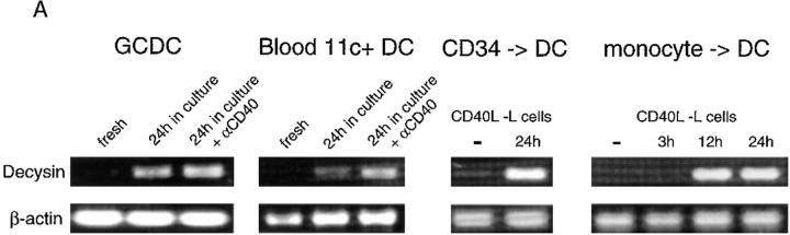
To identify genes expressed by a specific subset of dendritic cells found in vivo a polymerase chain reaction-based cDNA subtraction technique was applied to the recently described germinal center dendritic cells. A novel member of the disintegrin metalloproteinase family was cloned which comprises a not typical zinc-chelating catalytic site most similar to a bacterial metalloproteinase. Dendritic cell precursors or immature dendritic cells express no or low levels of the message. It is induced to high levels upon spontaneous or CD40-dependent maturation and in a mixed lymphocyte reaction. In situ hybridization showed distinct expression of this gene in the germinal center. This, together with the findings that certain disintegrin metalloproteinases regulate the activity of tumor necrosis factor alpha and that metalloproteinases have also been implicated in FasL processing, suggest that this novel molecule may play an important role in dendritic cell function and their interactions with germinal center T cells.


Figures

Figure 2
Figure 2
Restricted expression profile of decysin by RT-PCR. (A) Decysin is not detected in PMA/ionomycin-activated human cell lines TF1 (myeloid precursor cell), JURKAT (T cell), CHA, MRC5 (kidney epithelial and lung fibroblastic cells), and JY (B cell), but is expressed in PMA/ionomycin-activated stem cell–derived DCs. PCR with specific primers to decysin (Fig. 3 A) and β actin was performed on reverse transcribed RNA from the cell lines, stem cell– derived DCs harvested at days 6 and 12 of cell culture (6) as well as GCDC library subtracted (Fig. 1, lane 7) and nonsubtracted (Fig. 1, lane 5). In the subtracted library, β actin could not be amplified. (B) A low level of cDNA is seen in PBMCs, monocytes and B cells. GCDCs were activated for 24 h by αCD40 antibody G28-5 in complete medium. Peripheral blood mononuclear cells, monocytes, and T lymphocytes were obtained from human peripheral blood, and B lymphocytes from human tonsils.
Figure 3
Figure 3
A novel member of the disintegrin metalloproteinases. (A) Nucleotide and amino acid sequence. The underlined nucleotides represent the original 744-bp RsaI fragment cloned from the GCDC library. The full-length cDNA was cloned from stem cell–derived DCs. The potential polyadenylation site 17 bp upstream of the polyA tail is underlined. Primers used in RT-PCR are shown, which generate a 562-bp product. The first two methionine translation initiation codons are highlighted in bold, and cysteine 187 of the conserved cysteine-switch activation mechanism is circled. The putative furin cleavage site (RISR) and the zinc-binding consensus are boxed. These sequence data are available from EMBL/GenBank/DDBJ under the accession number Y13323. (B) Comparison of the zinc-binding sites of decysin, ScNP from Streptomyces caespitosus (20), and the five subfamilies of zinc endoproteases (38): disintegrin metalloproteinases (snake venom H2 proteinase [39]), matrix metalloproteinases (collagenase [40]), astacin (41), serralysin (42), and thermolysin (43). Decysin and ScNP are the only metalloproteinases in which an aspartic acid (D) replaces histidine (H) as the third zinc-chelating amino acid. (C) Alignment of the disintegrin domains of decysin, mammalian disintegrin metalloproteinases, and the snake disintegrin Ht-e. Decysin displays strong amino acid conservation along the disintegrin domain before prematurely terminating. Other protein sequences continue beyond the last cysteine. EMBL/GenBank/DDBJ accession numbers are given in the figure.
Figure 3
Figure 3
A novel member of the disintegrin metalloproteinases. (A) Nucleotide and amino acid sequence. The underlined nucleotides represent the original 744-bp RsaI fragment cloned from the GCDC library. The full-length cDNA was cloned from stem cell–derived DCs. The potential polyadenylation site 17 bp upstream of the polyA tail is underlined. Primers used in RT-PCR are shown, which generate a 562-bp product. The first two methionine translation initiation codons are highlighted in bold, and cysteine 187 of the conserved cysteine-switch activation mechanism is circled. The putative furin cleavage site (RISR) and the zinc-binding consensus are boxed. These sequence data are available from EMBL/GenBank/DDBJ under the accession number Y13323. (B) Comparison of the zinc-binding sites of decysin, ScNP from Streptomyces caespitosus (20), and the five subfamilies of zinc endoproteases (38): disintegrin metalloproteinases (snake venom H2 proteinase [39]), matrix metalloproteinases (collagenase [40]), astacin (41), serralysin (42), and thermolysin (43). Decysin and ScNP are the only metalloproteinases in which an aspartic acid (D) replaces histidine (H) as the third zinc-chelating amino acid. (C) Alignment of the disintegrin domains of decysin, mammalian disintegrin metalloproteinases, and the snake disintegrin Ht-e. Decysin displays strong amino acid conservation along the disintegrin domain before prematurely terminating. Other protein sequences continue beyond the last cysteine. EMBL/GenBank/DDBJ accession numbers are given in the figure.
Figure 1
Figure 1
Construction of a representative cDNA library from three million CD40-activated GCDCs, subtracted against U937 cDNA. (A) Tester cDNA was cut with RsaI, and adapters were ligated and amplified by adapter-specific (round 1) and adapter-nested (round 2) primers in the absence (lanes 1, 2 and 5, 6) or presence (lanes 3, 4 and 7, 8) of driver cDNA. As positive control skeletal muscle cDNA containing a trace amount of ΦX174 (HaeIII) marker DNA (0.02% of total cDNA) was subtracted against skeletal muscle cDNA (lane 4). (B) 10 times more subtracted GCDC cDNA than in lane 8 was resolved on a long-run low melting agarose gel. DNA within the bracket was recovered and cloned.
Figure 4
Figure 4
Decysin is highly expressed in mature DCs. (A) 28 cycle PCR on GCDCs and CD11c+ blood DCs immediately after cell sorting, after 24 h incubation in complete medium and after CD40 stimulation for 24 h in complete medium. Day 12 harvested CD34 stem cell– and monocyte-derived DC were analyzed for decysin before and after CD40 ligation on CD40L-transfected mouse L cells for the indicated time. (B) PCR on stem cell–generated DCs before and after coculture with alloreactive total naive T cells for 12 and 24 h. β actin was amplified with 28 cycles, decysin, CD83, and IL-2 with 35 cycles.
Figure 4
Figure 4
Decysin is highly expressed in mature DCs. (A) 28 cycle PCR on GCDCs and CD11c+ blood DCs immediately after cell sorting, after 24 h incubation in complete medium and after CD40 stimulation for 24 h in complete medium. Day 12 harvested CD34 stem cell– and monocyte-derived DC were analyzed for decysin before and after CD40 ligation on CD40L-transfected mouse L cells for the indicated time. (B) PCR on stem cell–generated DCs before and after coculture with alloreactive total naive T cells for 12 and 24 h. β actin was amplified with 28 cycles, decysin, CD83, and IL-2 with 35 cycles.
Figure 5
Figure 5
Northern analysis. PolyA+ RNA from different human tissues was hybridized with a specific decysin probe. Shown here are multiple tissue blot II (No. 7759-1; Clontech) and immune systems blot (No. 7754-1; Clontech). Not shown are tissue blots No. 7760-1 and No. 7756-1 as they were completely negative. Exposure time was 2 wk.
Figure 6
Figure 6
Localization of decysin mRNA to germinal centers by in situ hybridization. Human tonsil sections are hybridized with antisense (A, C) and sense (B) decysin 35S-labeled RNA probes. (A) and (B) are from serial sections. Original magnification: 40. The germinal center marked by an arrow original magnification: 100 (C). The sense probe generates diffuse background hybridization, whereas with the antisense probe, dense clusters of silver grains are seen mainly localized to the follicles. D is from reference , and shows immunohistological staining of CD4+CD11c+ GCDCs with anti-CD11c (red). In blue are proliferating cells stained by anti-Ki67. Original magnification: 100.

References

    1. Cella M, Sallusto F, Lanzavecchia A. Origin, maturation and antigen presenting function of dendritic cells. Curr Opin Immunol. 1997;9:10–16. - PubMed
    1. Caux, C., and J. Banchereau. 1996. In vitro regulation of dendritic cell development and function. In Blood Cell Biochemistry. T. Whetton and J. Gordon, editors. Plenum Press, London. 263–301.
    1. Austyn JD. New insights into the mobilization and phagocytic activity of dendritic cells. J Exp Med. 1996;183:1287–1292. - PMC - PubMed
    1. Grouard G, Durand I, Filgueira L, Banchereau J, Liu YJ. Dendritic cells capable of stimulating T cells in germinal centers. Nature (Lond) 1996;384:364–367. - PubMed
    1. Caux C, Dezutter-Dambuyant C, Schmitt D, Banchereau J. GM-CSF and TNF-α cooperate in the generation of dendritic Langerhans cells. Nature (Lond) 1992;360:258–261. - PubMed

MeSH terms

Associated data