. 1997 Aug 29;186(5):665-72.
doi: 10.1084/jem.186.5.665.

High levels of a major histocompatibility complex II-self peptide complex on dendritic cells from the T cell areas of lymph nodes

Affiliations

High levels of a major histocompatibility complex II-self peptide complex on dendritic cells from the T cell areas of lymph nodes

K Inaba et al. J Exp Med. .

Abstract

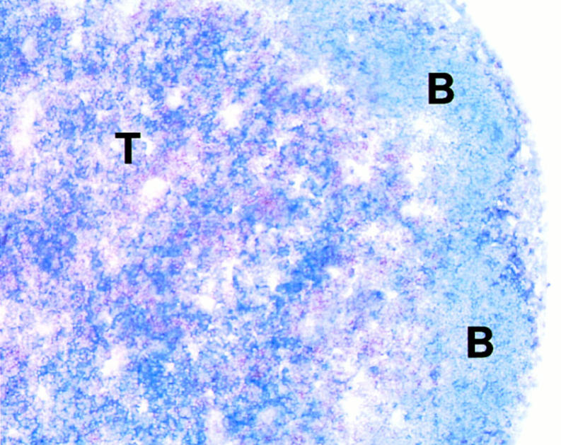
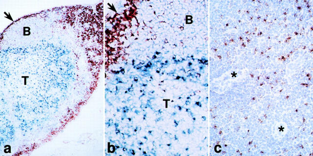
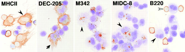
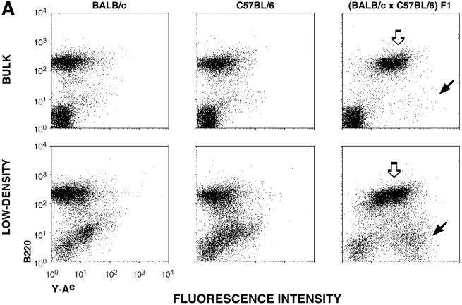
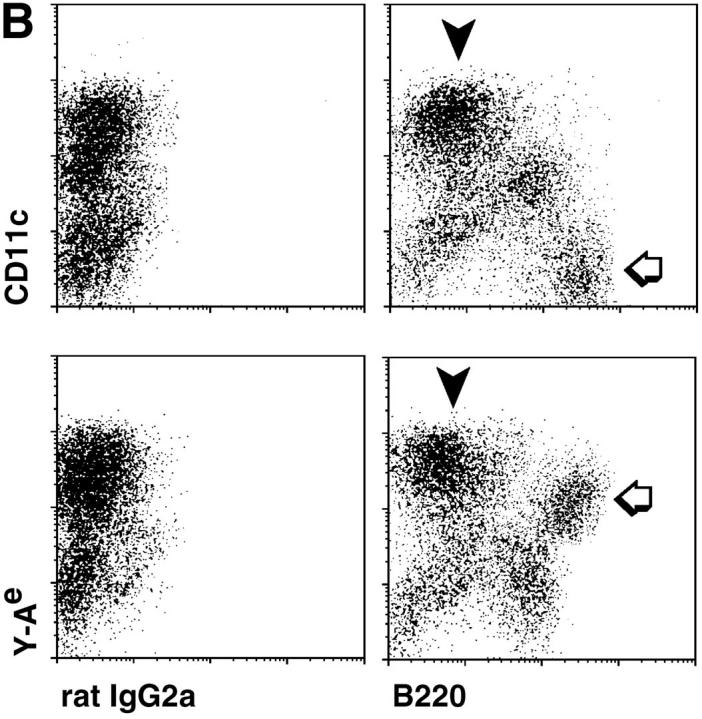
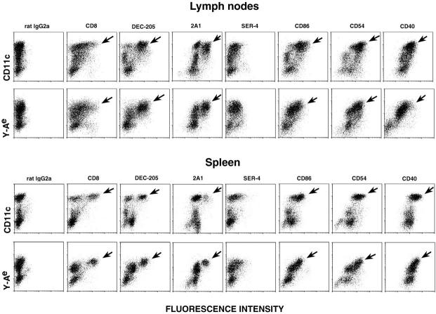
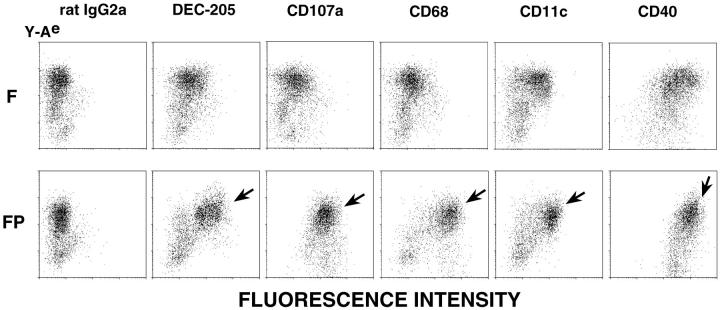
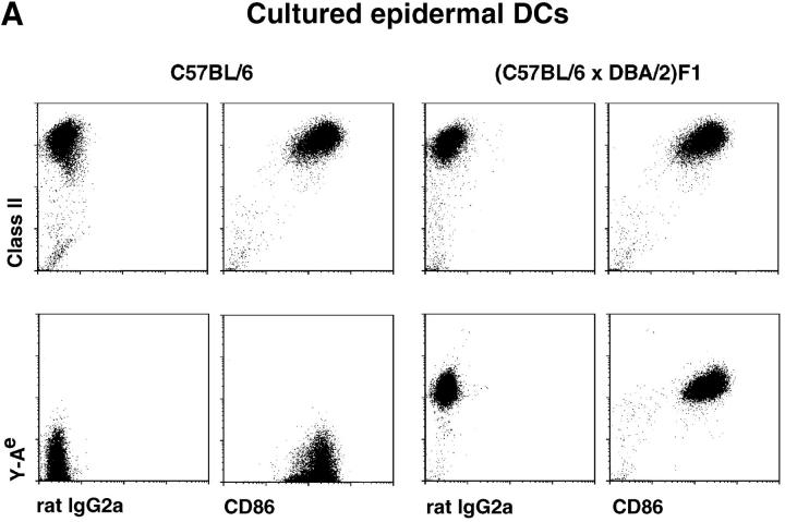
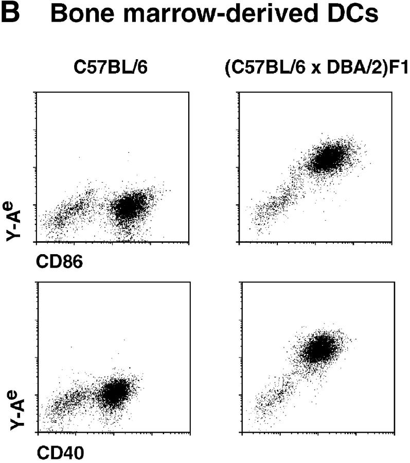
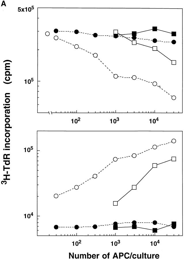
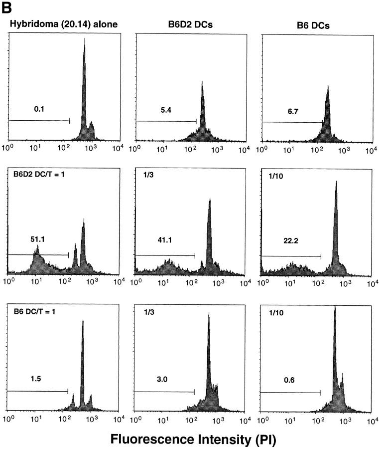
T lymphocytes recirculate continually through the T cell areas of peripheral lymph nodes. During each passage, the T cells survey the surface of large dendritic cells (DCs), also known as interdigitating cells. However, these DCs have been difficult to release from the lymph node. By emphasizing the use of calcium-free media, as shown by Vremec et al. (Vremec, D., M. Zorbas, R. Scollay, D.J. Saunders, C.F. Ardavin, L. Wu, and K. Shortman. 1992. J. Exp. Med. 176:47-58.), we have been able to release and enrich DCs from the T cell areas. The DCs express the CD11c leukocyte integrin, the DEC-205 multilectin receptor for antigen presentation, the intracellular granule antigens which are recognized by monoclonal antibodies M342, 2A1, and MIDC-8, very high levels of MHC I and MHC II, and abundant accessory molecules such as CD40, CD54, and CD86. When examined with the Y-Ae monoclonal which recognizes complexes formed between I-Ab and a peptide derived from I-Ealpha, the T cell area DCs expressed the highest levels. The enriched DCs also stimulated a T-T hybridoma specific for this MHC II-peptide complex, and the hybridoma underwent apoptosis. Therefore DCs within the T cell areas can be isolated. Because they present very high levels of self peptides, these DCs should be considered in the regulation of self reactivity in the periphery.


Figures

Figure 1
Figure 1
Stronger staining for MHC–self peptide complexes in lymph node T cell (T) vs. B cell areas (B). Double labeling for Y-Ae (blue) and CD8+ T cells (brown).
Figure 2
Figure 2
Staining for the 2A1 antigen which is abundant in DCs in lymph node T areas. (a and b) Low and high power views for macrophages (sialoadhesin+) in the subcapsular sinus (arrows) and medulla (brown) and the 2A1 antigen in the deep cortex (T area) and to a less extent B cell follicles (blue). (c) The deep cortex with several venules (*) is stained for the 2A1 antigen (brown) and counterstained with hematoxylin.
Figure 3
Figure 3
Enrichment of lymph node cells (see Materials and Methods) with many markers of T cell area DCs: MHC II, DEC-205, M342+ and MIDC-8+ granules, and B220+ B cells. Typical DCs (black arrowheads) and B cells (white arrowhead) are present.
Figure 4
Figure 4
Double labeling of lymph node, low density cells for CD11c integrin, B220 and Y-Ae. (A) In bulk lymph node suspensions, most of the Y-Ae+ cells are B220+ and only found in the appropriate mouse strain, BALB/c × C57BL/6 (white arrow), but by enriching DCs in a fraction with a low buoyant density, a B220 population with very high levels of Y-Ae emerges (black arrow). (B) Double labeling with B220 and either CD11c or Y-Ae to show the DCs (black arrowheads) and B cells (white arrows).
Figure 4
Figure 4
Double labeling of lymph node, low density cells for CD11c integrin, B220 and Y-Ae. (A) In bulk lymph node suspensions, most of the Y-Ae+ cells are B220+ and only found in the appropriate mouse strain, BALB/c × C57BL/6 (white arrow), but by enriching DCs in a fraction with a low buoyant density, a B220 population with very high levels of Y-Ae emerges (black arrow). (B) Double labeling with B220 and either CD11c or Y-Ae to show the DCs (black arrowheads) and B cells (white arrows).
Figure 5
Figure 5
Double labeling of lymph node and spleen for CD11c or Y-Ae on the y axis and a panel of mAbs on the x axis. All cells were fixed in formaldehyde and permeabilized with saponin to reveal predominantly intracellular antigens like DEC-205 and 2A1 (see Fig. 6). In lymph node, many CD11c+ cells and the strongest Y-Ae+ cells express the markers of T cell area DCs (black arrows). In spleen, only a subset of CD11c+ cells, but all the strong Y-Ae+ cells, carry markers of T cell area DCs (black arrows).
Figure 6
Figure 6
Markers of T cell area DCs are identified after saponin permeabilization. Low density, lymph node cells, fixed with HCHO (F) or fixed and permeabilized with saponin (FP), were stained with biotin Y-Ae (y axis) and mAbs (x axis). DEC-205, CD107a (LAMP-1), and CD68 are all found in the endocytic system.
Figure 7
Figure 7
DCs which have not been exposed to B cells express high levels of the Y–Ae, MHC–peptide complex. DCs were isolated from cultures which lack B cells, epidermal cells (A) and bone marrow stimulated with GM-CSF (B). In each instance, the DCs have high levels of CD86 and MHC II or MHC II–self peptide complexes.
Figure 7
Figure 7
DCs which have not been exposed to B cells express high levels of the Y–Ae, MHC–peptide complex. DCs were isolated from cultures which lack B cells, epidermal cells (A) and bone marrow stimulated with GM-CSF (B). In each instance, the DCs have high levels of CD86 and MHC II or MHC II–self peptide complexes.
Figure 8
Figure 8
Sorted lymph node DCs are potent APCs for IL-2 release and death of Y-Ae-specific, T-T hybridomas. Function is shown for sorted DCs and B cells from C57BL/6 and B6 × D2 F1 mice. (A) The response was measured as IL-2 release (bottom, proliferation of conA blasts) or 3H-TdR uptake (top) by the T-T hybrid, the latter being reduced by apoptosis. ▪, C57BL/6 B cells; □, B6D2F1 B cells; --•--, C57BL/6 DCs; --○-- B6D2F1 DCs; ○, None. (B) Apoptosis was measured by identifying hypodiploid profiles using propidium iodide staining. Extensive apoptosis occurs when the DCs are from B6D2 mice which express the Y-Ae epitope.
Figure 8
Figure 8
Sorted lymph node DCs are potent APCs for IL-2 release and death of Y-Ae-specific, T-T hybridomas. Function is shown for sorted DCs and B cells from C57BL/6 and B6 × D2 F1 mice. (A) The response was measured as IL-2 release (bottom, proliferation of conA blasts) or 3H-TdR uptake (top) by the T-T hybrid, the latter being reduced by apoptosis. ▪, C57BL/6 B cells; □, B6D2F1 B cells; --•--, C57BL/6 DCs; --○-- B6D2F1 DCs; ○, None. (B) Apoptosis was measured by identifying hypodiploid profiles using propidium iodide staining. Extensive apoptosis occurs when the DCs are from B6D2 mice which express the Y-Ae epitope.

References

    1. Liu Y-J, Grouard G, de Bouteiller O, Banchereau J. Follicular dendritic cells and germinal centers. Int Rev Cytol. 1996;166:139–179. - PubMed
    1. Szakal AK, Kosco MH, Tew JG. Microanatomy of lymphoid tissue during humoral immune responses: structure function relationships. Annu Rev Immunol. 1989;7:91–109. - PubMed
    1. Steinman RM. The dendritic cell system and its role in immunogenicity. Annu Rev Immunol. 1991;9:271–296. - PubMed
    1. Williams LA, Egner W, Hart DN. Isolation and function of human dendritic cells. Int Rev Cytol. 1994;153:41–103. - PubMed
    1. Steinman RM, Pack M, Inaba K. Dendritic cells in the T cell areas of lymphoid organs. Immunol Rev. 1997;156:25–37. - PubMed

Publication types

MeSH terms