. 1997 Aug 29;186(5):719-30.
doi: 10.1084/jem.186.5.719.

The lymphocyte function-associated antigen 1 I domain is a transient binding module for intercellular adhesion molecule (ICAM)-1 and ICAM-3 in hydrodynamic flow

Affiliations

The lymphocyte function-associated antigen 1 I domain is a transient binding module for intercellular adhesion molecule (ICAM)-1 and ICAM-3 in hydrodynamic flow

R Knorr et al. J Exp Med. .

Abstract

The I domain of lymphocyte function-associated antigen (LFA)-1 contains an intercellular adhesion molecule (ICAM)-1 and ICAM-3 binding site, but the relationship of this site to regulated adhesion is unknown. To study the adhesive properties of the LFA-1 I domain, we stably expressed a GPI-anchored form of this I domain (I-GPI) on the surface of baby hamster kidney cells. I-GPI cells bound soluble ICAM-1 (sICAM-1) with a low avidity and affinity. Flow cell experiments demonstrated a specific rolling interaction of I-GPI cells on bilayers containing purified full length ICAM-1 or ICAM-3. The LFA-1 activating antibody MEM-83, or its Fab fragment, decreased the rolling velocity of I-GPI cells on ICAM-1-containing membranes. In contrast, the interaction of I-GPI cells with ICAM-3 was blocked by MEM-83. Rolling of I-GPI cells was dependent on the presence of Mg2+. Mn2+ only partially substituted for Mg2+, giving rise to a small fraction of rolling cells and increased rolling velocity. This suggests that the I domain acts as a transient, Mg2+-dependent binding module that cooperates with another Mn2+-stimulated site in LFA-1 to give rise to the stable interaction of intact LFA-1 with ICAM-1.


Figures
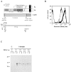
Figure 1
Figure 1
Generation of a glycolipid-anchored LFA-1 I domain (I-GPI). (A) Schematic of LFA-1 and I-GPI with PCR strategy. The signal peptide of the LFA-1 α chain was linked to the I domain with six amino acids of repeat II as a linker. An engineered HindIII site at the 3′ end of the coding region for the I domain was used to subclone the I domain containing PCR product in frame into a GPI-anchoring vector. (B) The expression of I-GPI on BHK cells was tested by FACScan® using TS1/22 IgG (bold solid), MEM-83 IgG (solid, superimposed with TS1/22 IgG), MEM-83 Fab (fine dotted), or TS2/4 IgG (fine solid, LFA-1 α, non–I domain) antibodies. (C) Western blot analysis of I-GPI cells treated with 0 (lanes 3 and 4), 0.1 (lanes 5 and 6), 0.2 (lanes 7 and 8) or 0.6 (lanes 9 and 10) U of PIPLC. Cells (p) and supernatant (s) were separated and subjected to SDS-PAGE. Supernatant corresponding to 1.5 × 10⁵ cells/lane after PIPLC treatment was loaded, whereas the pellet was diluted fourfold. BHK/ VP16 cells served as control. TS1/22 as primary antibody was used.
Figure 2
Figure 2
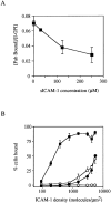
Competition of sICAM-1 with 125I TS1/22 Fab fragments for binding to I-GPI (A) and static adhesion assay of SKW3 cells and I-GPI cells to ICAM-1 coated on plates (B). (A) I-GPI E6 cells were incubated with the indicated concentrations of sICAM-1 and 125I TS1/22 Fab fragments (2 nM) for 20 min at room temperature. Cells and supernatant were separated by centrifugation through an oil cushion. The binding of TS1/22 Fab fragments is expressed as a fraction of I domain sites. Protein concentration in the assay was kept constant with ovalbumin. The result represents one experiment out of three. (B) Unstimulated SKW3 cells (open squares), SKW3 cells preincubated with 50 ng/ml PMA (filled squares), and I-GPI E6 cells (closed circles) or I-GPI E5 cells (open circles) were tested for adhesion to ICAM-1–coated plastic at 37°C in RPMI-1640, 2% BSA. Purified ICAM-1 was coated on polystyrene at the indicated density as determined by immunometric assay. SKW3 cells were labeled with calcein AM and BHK cells with BCECF.
Figure 3
Figure 3
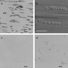
Stroboscopic images of free, attached, and rolling cells. 16 images spanning 6 s digitized from S-VHS tape (a, c, and d) or 8 images spanning 4 s directly acquired with a cooled CCD camera (40-ms exposure) (b) were added together such that the motion of cells in the flow field over time is represented in a single image. (a) I-GPI–transfected cells; (b) same as a at a higher magnification; (c) vector transfected cells; (d) ICAM-1 GPI-transfected cells. The bilayer membranes contained 500 molecules/μm2 ICAM-1 (a–c) or 500 molecules/μm2 LFA-1 (d). Scale bars = 50 μm.
Figure 4
Figure 4
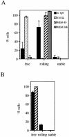
Effect of different anti–LFA-1 I domain antibodies on the interaction between I-GPI E6 cells and ICAM-1 (A) or ICAM-3 (B) containing bilayers at a flow rate of 1.1 dyn/cm2. I-GPI E6 cells in HBS/BSA buffer containing 2 mM MgCl2 were not treated (black) or pretreated with the antibodies TS1/22, MEM-83, or MEM-83 Fab fragments, and tested for rolling adhesion on bilayers at 500 ICAM-1 molecules/μm2 (A) or bilayers at 1,000 ICAM-3 molecules/μm2 (B). Error bars represent 95% confidence interval on the mean of free, rolling, or attached cells. P <0.03 for rolling cells on ICAM-3.
Figure 5
Figure 5
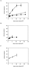
Rolling interaction of I-GPI cells with ICAM-1– (A and B) or ICAM-3– (C) containing bilayers. The rolling velocity of I-GPI E6 cells treated with no IgG (solid square), MEM-83 IgG (solid circle), or MEM-83 Fab (open circle) on membranes containing 500 molecules/μm2 (A) or 150 molecules/μm2 ICAM-1 (B) or 1,000 molecules/μm2 ICAM-3 (C) as function of shear stress is shown. Error bars represent 95% confidence interval on the mean rolling velocity.
Figure 6
Figure 6
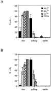
Ion dependency of the interaction of I-GPI E6 cells with ICAM-1–containing membranes in the absence (A) and presence (B) of MEM-83. I-GPI E6 cells in HBS/BSA buffer containing 2 mM MgCl2 (black), 0.2 mM MnCl2 (stipled), 1 mM CaCl2 (diagonal stripes), or 2 mM EDTA (vertical stripes) were not treated (A) or pretreated with MEM-83 (0.1 mg/ml) (B), washed and assayed on bilayer membranes with 500 ICAM-1 molecules/μm2 at a flow rate of 1.1 dyn/cm2. Error bars represent 95% confidence interval on the mean of free, rolling, or attached cells.
Figure 7
Figure 7
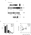
The distance of the I domain from the bilayer membrane does not result in a stable interaction. (A) Schematic of I-EF and I-CD2. In I-EF, the I domain was extended with repeats III, IV, V, VI, and the first few amino acids of repeat VII of the LFA-1 α chain. I-EF is linked to the Decay Accelerating Factor– GPI-anchoring signal as in I-GPI. In I-CD2, the spacer between the I domain and the Decay Accelerating Factor–GPI-anchoring signal consists of a few amino acids of the domain 1, the linker region, domain 2, and the stalk of human CD2. (B) Interaction of COS-1 cells transiently transfected with I-GPI, I-CD2, or I-EF, on membranes containing 500 molecules/μm2 ICAM-1 in HBS/BSA containing 2 mM MgCl2. The data were normalized according to a FACS® analysis and are presented as percent transfectants. The rolling interaction was statistically significant with P <0.01 for each construct. (C) The rolling velocity of COS-1 cells transiently transfected with I-GPI, I-CD2, or I-EF on membranes containing 500 molecules/μm2 ICAM-1 in HBS/BSA buffer containing 2 mM MgCl2 was determined.

References

    1. Dustin ML, Springer TA. Role of lymphocyte adhesion receptors in transient interactions and cell locomotion. Annu Rev Immunol. 1991;9:27–66. - PubMed
    1. Lub M, van Kooyk Y, Figdor CG. Ins and outs of LFA-1. Immunol Today. 1995;16:479–483. - PubMed
    1. Stewart MP, Hogg N. Regulation of leukocyte integrin function: affinity vs. avidity. J Cell Biochem. 1996;61:554–561. - PubMed
    1. Stanley P, Bates PA, Harvey J, Bennett RI, Hogg N. Integrin LFA-1 α subunit contains an ICAM-1 binding site in domains V and VI. EMBO (Eur Mol Biol Organ) J. 1994;13:1790–1798. - PMC - PubMed
    1. Goodman TG, Bajt ML. Identifying the putative metal ion-dependent adhesion site in the β2 (CD18) subunit required for αLβ2 and αMβ2ligand interactions. J Biol Chem. 1996;271:23729–23736. - PubMed

Publication types

MeSH terms