. 1997 Sep 2;94(18):9585-9.
doi: 10.1073/pnas.94.18.9585.

The yeast peptide-methionine sulfoxide reductase functions as an antioxidant in vivo

Affiliations

The yeast peptide-methionine sulfoxide reductase functions as an antioxidant in vivo

J Moskovitz et al. Proc Natl Acad Sci U S A. .

Abstract

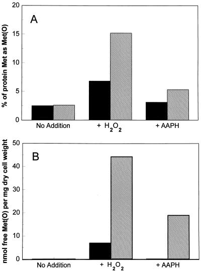
A gene homologous to methionine sulfoxide reductase (msrA) was identified as the predicted ORF (cosmid 9379) in chromosome V of Saccharomyces cerevisiae encoding a protein of 184 amino acids. The corresponding protein has been expressed in Escherichia coli and purified to homogeneity. The recombinant yeast MsrA possessed the same substrate specificity as the other known MsrA enzymes from mammalian and bacterial cells. Interruption of the yeast gene resulted in a null mutant, DeltamsrA::URA3 strain, which totally lost its cellular MsrA activity and was shown to be more sensitive to oxidative stress in comparison to its wild-type parent strain. Furthermore, high levels of free and protein-bound methionine sulfoxide were detected in extracts of msrA mutant cells relative to their wild-type parent cells, under various oxidative stresses. These findings show that MsrA is responsible for the reduction of methionine sulfoxide in vivo as well as in vitro in eukaryotic cells. Also, the results support the proposition that MsrA possess an antioxidant function. The ability of MsrA to repair oxidative damage in vivo may be of singular importance if methionine residues serve as antioxidants.


Figures

Figure 2
Figure 2
Interruption of the msrA gene in yeast cells. Haploid strains H8 and H9 were transformed with a DNA fragment containing the disrupted msrA gene by the URA3 gene insertion. The cells were grown on minimal medium plates without uracil. Several colonies were collected and grown on minimal liquid medium without uracil. DNA was isolated from the different transformants, and the presence of disrupted msrA gene was assayed by PCR. PCR products are shown in the agarose gel, using oligonucleotides H1 and H2, of the wild-type and the disruptant strains and the original DNA construct used for the disruption of the gene. Lanes: 1, DNA fragment used for the interruption of the gene; 2 and 4, PCR product of DNA isolated from H8 and H9 wild-type cells, respectively; 3 and 5, PCR product of DNA isolated from msrA disruptant cells of H8 and H9 strains, respectively.
Figure 1
Figure 1
SDS/PAGE analysis of fractions during the purification of recombinant yeast MsrA protein. M15 cells were transformed with pQE-30 harboring the yeast msrA gene and the expressed protein was purified as described. Lanes: 1, S-30 − IPTG; 2, S-30 + IPTG; 3, yeast MsrA protein eluted from the Ni-NTA resin after treatment with buffer A containing 400 mM imidazole. The arrow indicates where MsrA migrates.
Figure 3
Figure 3
Growth of yeast strains in the presence and absence of H2O2. Yeast from stationary phase cultures were inoculated into yeast synthetic medium at 1:300 dilution and were grown aerobically at 30°C with or without H2O2 (1 mM). The symbols are defined as follows: wild-type parent strain (H9) grown with (▪) or without (□) H2O2; and H9 ΔmsrA::URA3 strain grown with (•) or without (○) H2O2.
Figure 4
Figure 4
Met(O) content in yeast strains that were grown under different oxidative stress conditions. Each yeast strain was generally grown in a synthetic medium as described in Fig. 3 until the cell density reached 300 Klett units under each culture condition. Then cells were harvested and their extracts were measured for protein-bound Met(O) (A) or free Met(O) (B), as described. Filled bars represent wild-type (H9) strain and hatched bars represent H9 ΔmsrA::URA3 strain. Similar results were obtained with the H8 yeast strain and its corresponding null msrA mutant strain. H2O2 and AAPH concentrations were 1 mM and 6 mM, respectively.

References

    1. Moskovitz J, Weissbach H, Brot N. Proc Natl Acad Sci USA. 1995;93:2095–2099. - PMC - PubMed
    1. Moskovitz J, Jenkins N A, Gilbert D J, Copeland N G, Jursky F, Weissbach H, Brot N. Proc Natl Acad Sci USA. 1995;93:3205–3208. - PMC - PubMed
    1. Brot N, Fliss H, Coleman T, Weissbach H. Methods Enzymol. 1984;107:352–360. - PubMed
    1. Babior B M. N Engl J Med. 1978;298:659–669. - PubMed
    1. Moskovitz J, Rahman A M, Strassman J, Yancey S O, Kushner S R, Brot N, Weissbach H. J Bacteriol. 1995;177:502–507. - PMC - PubMed

MeSH terms