. 1997 Sep 2;94(18):9920-5.
doi: 10.1073/pnas.94.18.9920.

Inactivation of the survival motor neuron gene, a candidate gene for human spinal muscular atrophy, leads to massive cell death in early mouse embryos

Affiliations

Inactivation of the survival motor neuron gene, a candidate gene for human spinal muscular atrophy, leads to massive cell death in early mouse embryos

B Schrank et al. Proc Natl Acad Sci U S A. .

Abstract

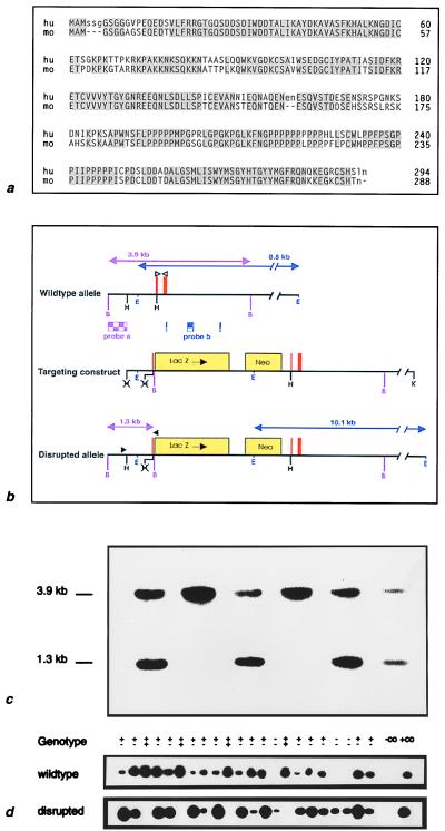
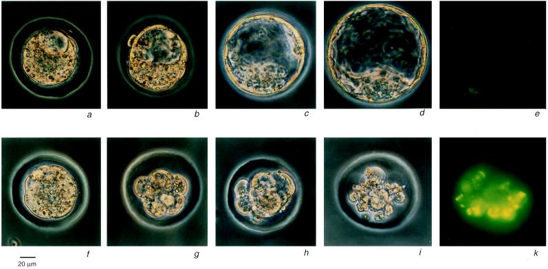
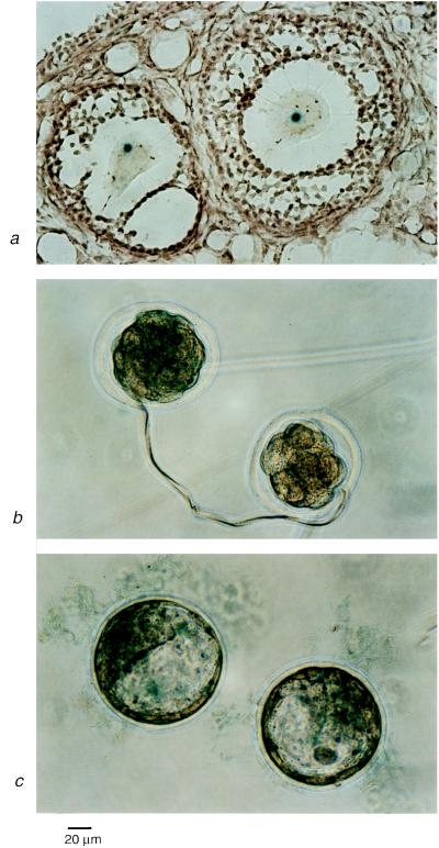
Proximal spinal muscular atrophy is an autosomal recessive human disease of spinal motor neurons leading to muscular weakness with onset predominantly in infancy and childhood. With an estimated heterozygote frequency of 1/40 it is the most common monogenic disorder lethal to infants; milder forms represent the second most common pediatric neuromuscular disorder. Two candidate genes-survival motor neuron (SMN) and neuronal apoptosis inhibitory protein have been identified on chromosome 5q13 by positional cloning. However, the functional impact of these genes and the mechanism leading to a degeneration of motor neurons remain to be defined. To analyze the role of the SMN gene product in vivo we generated SMN-deficient mice. In contrast to the human genome, which contains two copies, the mouse genome contains only one SMN gene. Mice with homozygous SMN disruption display massive cell death during early embryonic development, indicating that the SMN gene product is necessary for cellular survival and function.


Figures

Figure 1
Figure 1
Disruption of the murine SMN gene. (a) Amino acid sequence of the human (10) and mouse SMN protein. Uppercase letters represent related residues, shaded boxes highlight identities. The protein appears highly conserved with 83% identical and 98% related residues (31). The largest uninterrupted region of complete identity is found near the amino terminus with 46 aa. The characteristic proline-rich region from aa 190–246 (54% proline content) is also preserved. The expected Mr is 31.2 kDa. (b) Targeted disruption of the SMN gene. Partial genomic structure of the mouse SMN locus. Sizes of diagnostic restriction fragments are shown as arrows (purple, 5′ end; blue, 3′ end). Tiled boxes represent the DNA probes used for confirmation of targeting events. Solid triangles indicate primer position for the PCR screen of the mutated locus, open triangles indicate primer position for amplification of DNA corresponding to the wild-type allele. The relative position of exon 2 is shown by red bars. While splice junctions in the coding region are generally preserved between human and mouse, exon 2 of the mouse gene is fragmented by a small 200 bp intron. The HindIII site in exon 2 (aa 38 and 39) was used to disrupt the gene by insertion of the targeting vector pGNA (25) providing an in-frame lacZ fusion and the neor gene under a separate promoter. Genes supplied by pGNA are shown as yellow boxes (not drawn to scale). B, BamHI; H, HindIII; E, EcoRI; K, KpnI. (c) Genotyping of heterozygous intercross progeny: Densitometric analysis of signal intensities of bands corresponding to mutated and wild-type alleles revealed a ratio of 1.1 ± 0.15 (SD, n = 11). This confirms that the SMN gene is not duplicated in mice. (d) PCR genotyping of blastocyst stage embryos. The wild-type allele was identified with primers corresponding to the 5′ and 3′ end of exon 2 (Upper). Primers shown in b were used to amplify the mutant allele (Lower).
Figure 2
Figure 2
Morphological alterations and TUNEL staining in control and mutant SMN embryos. (ae) Wild-type or heterozygous embryos. (fk) Homozygous mutant embryos at equivalent time points. A failure in transition to the blastocyst stage first became apparent at 80–90 hr after mating (g and h), with subsequent formation of a disorganized, multicystic structure (h) and finally extensive cellular degeneration (i) at 90–104 hr p.c. TUNEL staining of SMN−/− embryos showed strong staining of most cells, suggesting apoptotic cell death (k). In contrast, wild-type blastocysts showed only very few labeled cells (e).
Figure 3
Figure 3
SMN+/− lacZ Expression. (a) Strong expression was found in oocytes of pregnant mare serum gonadotrophin-treated female heterozygotes. (b and c) Embryo lacZ expression. Heterozygous males were mated to wild-type females to determine the onset of embryonic SMN expression. Whereas 5–8 cell morulae did not show any staining (b Right), compacted morulae (8–16 cells) expressed the reporter gene (b Left). β-Galactosidase activity was apparent in both inner cell mass and trophoblast cells at the blastocyst stage (c).

References

    1. Dubowitz V. Muscle Disorders in Childhood. London: Saunders; 1995. pp. 325–369.
    1. Pearn J H. Lancet. 1980;i:919–922. - PubMed
    1. Crawford T O. Neurology. 1996;46:335–340. - PubMed
    1. Munsat T L. Neuromuscular Disorders. 1991;1:81.
    1. Zerres K, Rudnik-Schöneborn S. Arch Neurol. 1995;52:518–523. - PubMed

Publication types

Substances

Associated data