. 1997 Sep 2;94(18):9956-61.
doi: 10.1073/pnas.94.18.9956.

Spinophilin, a novel protein phosphatase 1 binding protein localized to dendritic spines

Affiliations

Spinophilin, a novel protein phosphatase 1 binding protein localized to dendritic spines

P B Allen et al. Proc Natl Acad Sci U S A. .

Abstract

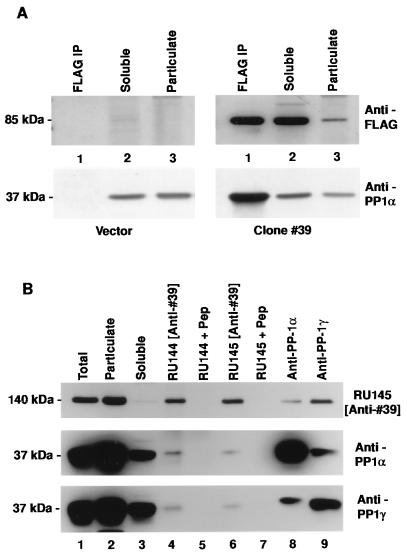
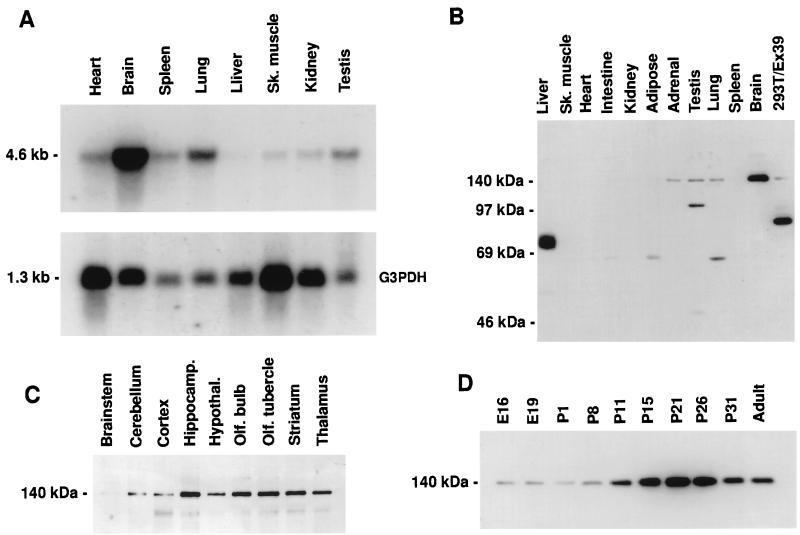
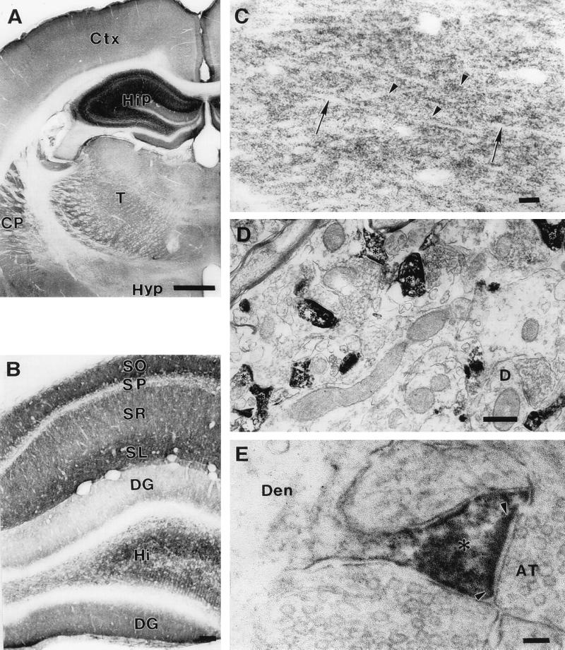
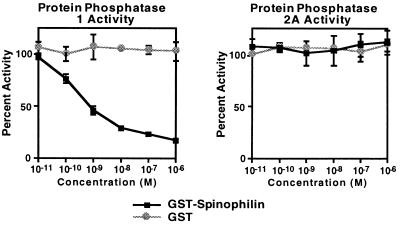
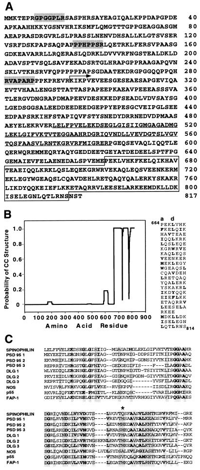
Dendritic spines receive the vast majority of excitatory synaptic contacts in the mammalian brain and are presumed to contain machinery for the integration of various signal transduction pathways. Protein phosphatase 1 (PP1) is greatly enriched in dendritic spines and has been implicated in both the regulation of ionic conductances and long-term synaptic plasticity. The molecular mechanism whereby PP1 is localized to spines is unknown. We have now characterized a novel protein that forms a complex with the catalytic subunit of PP1 and is a potent modulator of PP1 enzymatic activity in vitro. Within the brain this protein displays a remarkably distinct localization to the heads of dendritic spines and has therefore been named spinophilin. Spinophilin has the properties expected of a scaffolding protein localized to the cell membrane and contains a single consensus sequence in PSD95/DLG/zo-1, which implies cross-linking of PP1 to transmembrane protein complexes. We propose that spinophilin represents a novel targeting subunit for PP1, which directs the enzyme to those substrates in the dendritic spine compartment, e.g., neurotransmitter receptors, which mediate the regulation of synaptic function by PP1.


Figures

Figure 1
Figure 1
Clone #39 represents a PP1 binding protein. (A) Immunoblot analysis of proteins prepared from 293T cells transfected with expression vector alone (Left) or with clone #39 expression construct (Right). Lanes: 1, anti-FLAG immunoprecipitate; 2, 50 μg soluble fraction of cell lysate; 3, 50 μg particulate fraction of cell lysate. Blots were probed with anti-FLAG antibody (Upper), stripped, and reprobed with anti-PP1α antiserum (Lower). (B) Immunoblot analysis of proteins prepared from rat brain homogenate. Lanes: 1–3, 50 μg total lysate, 50 μg particulate fraction, 50 μg soluble fraction, respectively; 4–9, immunoprecipitates prepared from the soluble fraction using anti-clone #39 peptide antisera (RU144 and RU145 +/− immunogen peptide) or anti-PP1 antisera. The blot was probed sequentially with anti-clone #39 antiserum (Top), anti-PP1α antiserum (Middle), and anti-PP1γ antiserum (Bottom).
Figure 2
Figure 2
Expression pattern of mRNA and distribution of the endogenous protein represented by clone #39. (A) Northern blot analysis. (Upper) Detection of a transcript of ≈4.6 kb using a clone #39 probe. (Lower) Levels of the transcript for G3PDH to indicate relative mRNA loading. (B–D) Immunoblot analysis using anti-clone #39 antiserum RU145 and 50 μg per lane rat tissue homogenate in 1% SDS. (B) Expression of a protein migrating at ≈140 kDa in various rat tissues. Far right lane, 20 μg of 1% SDS homogenate from 293T cells expressing partial cDNA clone #39. Proteins detected by RU145 migrating at less than 140 kDa were not reactive with RU144 and vice versa (data not shown). These are presumably cross-reactive species. (C) Expression levels of the ≈140 kDa protein in various adult brain structures. (D) Developmental profile of the ≈140 kDa protein present in cortical homogenates at the embryonic (E) and postnatal (P) days indicated.
Figure 3
Figure 3
Neuronal distribution of spinophilin. (A) Light microscopic photomicrograph of a coronal section at the level of the hippocampus and thalamus. Immunoreactivity is most intense in the hippocampus (Hip). Moderate immunoreactivity is also present in the caudatoputamen (CP), thalamus (T), and cerebral cortex (Ctx). Immunoreactivity in the hypothalamus (Hyp) is light. (Bar = 1 mm.) (B) Light microscopic photomicrograph of the laminar pattern of immunoreactivity in a coronal section of the hippocampal formation. Immunoreactivity is most intense in stratum oriens (SO), stratum lacunosum moleculare (SL), and hilus (Hi). Immunoreactivity is moderately strong in stratum radiatum (SR) and absent from stratum pyramidale (SP). The upper, or ectal, limb of the dentate gyrus (DG) shows weaker immunoreactivity than the lower, or endal, limb. (Bar = 100 μm.) (C) High power light microscopic photomicrograph of the stratum radiatum showing immunoreactive puncta (arrowheads). Dendritic shafts (arrows) are not immunoreactive. (Bar = 10 μm.) (D) Electron micrograph showing immunolabeled dendritic spines (asterisks). An example of an unlabeled dendritic shaft that receives a synaptic contact in the plane of section is shown (D). (Bar = 500 nm.) (E) Electron micrograph showing immunoreactivity in a dendritic spine neck and especially in a spine head (asterisk). Immunoreactivity (black flocculent material) lines the cytoplasmic surface of the spine plasmalemma and coats the postsynaptic density (delineated by arrowheads). Immunoreactivity is not present in axon terminals (AT) or in the dendritic shaft (Den). (Bar = 100 nm.)
Figure 4
Figure 4
Effect of bacterial spinophilin fusion protein on the catalytic activity of PP1 and PP2A. Results (means ± SEM) were calculated as percent of activity in the absence of added bacterial protein.
Figure 5
Figure 5
Sequence characteristics of spinophilin. (A) Predicted amino acid sequence of spinophilin. The region encoded by the cDNA isolated in the library screen is indicated (arrow). Shaded heptamers indicate potential Src homology 3-binding domain motifs present in the proline-rich amino terminus. Underlined sequence indicates the position of a PDZ domain. Boxed sequence indicates a region predicted to form a coiled–coil structure. (B Left) Coiled–coil prediction. The mtidk matrix algorithm was used without weighting at the a and d positions (14). (Right) Alignment of amino acids in the C terminus of spinophilin as a heptad repeat. Hydrophobic residues are in boldface type and a stagger is introduced at the point where the prediction breaks down. (C) Amino acid alignment of the PDZ domain (492–583) of spinophilin with selected examples of other PDZ domain-containing proteins: PSD95 1(61–151)/2(156–246)/3(309–393), DLG 1(36–126)/2(150–244)/3(482–566), NOS (14–100), p55 (67–153), FAP-1 (86–179). Asterisk marks the first residue in the αB helix of the PDZ domain.

References

    1. Harris K M, Kater S B. Annu Rev Neurosci. 1994;17:341–371. - PubMed
    1. Ouimet C C, da Cruz e Silva E F, Greengard P. Proc Natl Acad Sci USA. 1995;92:3396–3400. - PMC - PubMed
    1. Surmeier D J, Bargas J, Hemmings H C, Jr, Nairn A C, Greengard P. Neuron. 1995;14:385–397. - PubMed
    1. Endo S, Critz S D, Byrne J H, Shenolikar S. J Neurochem. 1995;64:1833–1840. - PubMed
    1. Mulkey R M, Endo S, Shenolikar S, Malenka R C. Nature (London) 1994;369:486–488. - PubMed

Publication types

Associated data